Fisheries and Aquatic Sciences
BioMed Central
Research Article

A survey of epiphytic organisms in cultured kelp Saccharina japonica in Korea

Jong-Oh Kim1, Wi-Sik Kim1, Ha-Na Jeong1, Sung-Je Choi2, Jung-Soo Seo3, Myoung-Ae Park3, Myung-Joo Oh1,*
1Department of Aqualife MedicineJeonnam National University59626YeosuSouth Korea
2Algae Research Institute59146JeollaNamdoWandoSouth Korea
3Aquatic Life Disease Control Division, Fundamental Research DepartmentNational Fisheries Research and Development Institute46083BusanSouth Korea

© The Author(s) 2017. Open AccessThis article is distributed under the terms of the Creative Commons Attribution 4.0 International License (http://creativecommons.org/licenses/by/4.0/), which permits unrestricted use, distribution, and reproduction in any medium, provided you give appropriate credit to the original author(s) and the source, provide a link to the Creative Commons license, and indicate if changes were made. The Creative Commons Public Domain Dedication waiver (http://creativecommons.org/publicdomain/zero/1.0/) applies to the data made available in this article, unless otherwise stated.

Received: Aug 29, 2016; Accepted: Jan 10, 2017

Published Online: Jan 31, 2017

Abstract

A survey was conducted to investigate the presence of epiphytic organisms in four kelp Saccharina japonica farms in the coastal area of Korea from 2014 to 2015. Of 740 kelp samples that were taken, 208 exhibited six kinds of epiphytic organisms, including hydroid (detection rate: 11.6%), bryozoan (6.4%), polychaete (3.4%), algae (3.2%), caprellid (3%), and oyster (0.5%). The infestation rate for hydroid, bryozoan, and polychaete was significantly higher in the Wando farm, Busan farm, and Pohang farm, respectively. Epiphytic organisms were generally observed during May to September and not January to April, indicating that their infestation was significantly higher when the water had a higher temperature. The histopathogical examination revealed that hydroid and bryozoan organisms were attached on the cuticula of the thallus while some algae were attached on the cuticula of the thallus or had penetrated the epidermis. These results indicate that hydroid and bryozoan were the most predominant epiphytic organisms in Korean kelp farms, even though the infested thallus had not been broken.

Keywords: Epiphytic organisms; Bryozoan; Hydroid; Kelp; Saccharina japonica

Background

Within the last 30 years, seaweed cultivation has attained large-scale production levels and has become a major industry in China, Korea, and Japan. Korea had a harvest of 1,022,326 tons in 2012, which makes it the fourth largest seaweed-producing country in the world (FAO, 2014). The economically important seaweed species harvested in Korea are laver Porphyra spp., wakame Undaria spp., kelp Laminaria spp., fusiforme, Hizikia fusiformis, and green laver Monostroma spp. (KOSIS, 2015).

Kelp Saccharina japonica cultivation was industrialized in Korea in the 1970s, and from 2005, it has been extensively developed together with the abalone industry. During the 1970s to 1980s, kelp production reached less than 12,000 tons, and production steadily increased since then with about 370,000 tons harvested in 2013 (KOSIS, 2015). Kelp production now accounts for about 25% of all Korean seaweed cultivation (KOSIS, 2015). However, the increase in production has also resulted in kelp farms becoming severely infected by several epiphytic organisms (Gong et al., 2010; Park and Hwang, 2012). An infestation is characterized by the appearance of numerous colonies on the thallus of the kelp, and therefore, the affected kelp appears unsightly and cannot be sold. Although systematic research on fish disease has been conducted in Korea since the 1990s, seaweed disease has not yet been systematically studied. Therefore, little information is available on kelp disease. The present study consists of a survey that was conducted to investigate the presence of epiphytic organisms in Korean kelp farms. In addition, a histopathological examination was conducted to understand the effect by epiphytic organisms on the thallus.

Methods

Kelp samples and epiphytic organism examination

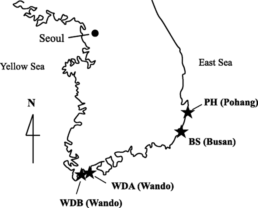
Kelp samples were obtained from four private farms in Wando (Southern Sea), Busan (Southern Sea), and Pohang (Eastern Sea) during 2014 and 2015 (Table 1, Fig. 1). Wando is the most popular area for kelp cultivation, producing about 97% of the kelp crop of Korea. The kelps were randomly selected by a farmer, transported on ice, and immediately subjected an examination of the epiphytic organisms. The total length (from apex to holdfast) of the thallus was measured, and the surfaces of the specimens were examined macroscopically and microscopically for the presence of epiphytic organisms. The detection rate of the epiphytic organisms was measured by calculating the percentages of the kelp infested by epiphytic organisms, and their infestation area was calculated as the percentage of the thallus that had been colonized. The number of caprellid was measured in a 2 × 2 cm kelp area infected with hydroid because their infestation area cannot be calculated. The epiphytic organisms that were obtained were photographed and identified according to their morphological characteristics (Gong et al., 2010; Kim and Lee, 1975; Lee, 1994; Park, 2010; 2011; Saunders and Metaxas, 2009; Schwaninger, 1999; The Korean Society of Systematic Zoology, 2014).

Table 1. Detection of epiphytic organisms in four kelp farms in Korea

Place

Farm

Date

Water temperature (°C)

Mean thallus length (cm)

Detection rate of epiphytic organisms % (detection no./total no.)

Hydroid

Bryozoan

Polychaete

Algae

Caprellid

Oyster

Wando

WDA

Apr 2014

11.5

340 (200–380)

0 (0/10)

0 (0/10)

0 (0/10)

0 (0/10)

0 (0/10)

0 (0/10)

May 2014

15.5

260 (260–270)

0 (0/2)

0 (0/2)

0 (0/2)

0 (0/2)

0 (0/2)

0 (0/2)

Jun 2014

17.4

NT

20 (1/5)

0 (0/5)

0 (0/5)

0 (0/5)

20 (1/5)

0 (0/5)

Jul 2014

21.7

190 (100–240)

100 (6/6)

50 (3/6)

0 (0/6)

100 (6/6)

0 (0/6)

0 (0/6)

Aug 2014

23

200 (100–240)

71.4 (5/7)

57.1 (4/7)

0 (0/7)

71.4 (5/7)

71.4 (5/7)

0 (0/7)

Sep 2014

22.1

120 (90–160)

57.1 (4/7)

71.4 (5/7)

0 (0/7)

14.3 (1/7)

0 (0/7)

57.1 (4/7)

Jan 2015

9

41 (20–71)

0 (0/71)

0 (0/71)

0 (0/71)

0 (0/71)

0 (0/71)

0 (0/71)

Jan 2015

8.3

82 (39–122)

0 (0/75)

0 (0/75)

0 (0/75)

0 (0/75)

0 (0/75)

0 (0/75)

Apr 2015

11

236 (200–280)

0 (0/30)

0 (0/30)

0 (0/30)

0 (0/30)

0 (0/30)

0 (0/30)

Total detection rate in WDA

7.5 (16/213)

5.6 (12/213)

0 (0/213)

5.6 (12/213)

2.8 (6/213)

0.9 (4/213)

WDB

May 2014

15.4

220 (100–330)

0 (0/14)

0 (0/14)

0 (0/14)

0(0/14)

0 (0/14)

0 (0/14)

Jun 2014

21

260 (180–320)

57.1 (4/7)

0 (0/7)

0 (0/7)

28.6 (2/7)

0 (0/7)

0 (0/7)

Jul 2014

21.5

160 (130–200)

100 (14/14)

0 (0/14)

35.7 (5/14)

0 (0/14)

7.1 (1/14)

0 (0/14)

Sep 2014

21.6

130 (60–230)

100 (15/15)

33.3 (5/15)

0 (0/15)

13.3 (2/15)

0 (0/15)

0 (0/15)

Feb 2015

9

57 (30–100)

0 (0/100)

0 (0/100)

0 (0/100)

0 (0/100)

0 (0/100)

0 (0/100)

Mar 2015

11

97 (50–180)

0 (0/73)

0 (0/73)

0 (0/73)

0 (0/73)

0 (0/73)

0 (0/73)

Apr 2015

12.5

127 (90–190)

0 (0/30)

0 (0/30)

0 (0/30)

0 (0/30)

0 (0/30)

0 (0/30)

Total detection rate in WDB

13 (33/253)

2 (5/253)

2 (5/253)

1.6 (4/253)

0.4 (1/253)

0 (0/253)

Busan

BS

Apr 2014

11

170 (50–270)

0 (0/10)

10 (1/10)

0 (0/10)

30 (3/10)

0 (0/10)

0 (0/10)

May 2014

14.5

230 (130–280)

100 (9/9)

88.9 (8/9)

0 (0/9)

0 (0/9)

0 (0/9)

0 (0/9)

Jun 2014

21

220 (180–300)

100 (7/7)

71.4 (5/7)

0 (0/7)

71.4 (5/7)

0 (0/7)

0 (0/7)

Jul 2014

20.4

160 (120–180)

100 (10/10)

90 (9/10)

0 (0/10)

0 (0/10)

100 (10/10)

0 (0/10)

Feb 2015

11.5

76 (26–175)

0 (0/58)

0 (0/58)

0 (0/58)

0 (0/58)

0 (0/58)

0 (0/58)

Feb 2015

12

143 (80–219)

0 (0/37)

0 (0/37)

0 (0/37)

0 (0/37)

0 (0/37)

0 (0/37)

Mar 2015

12.5

182 (110–260)

0 (0/70)

0 (0/70)

0 (0/70)

0 (0/70)

0 (0/70)

0 (0/70)

Apr 2015

13

203 (120–270)

0 (0/35)

0 (0/35)

0 (0/35)

0 (0/35)

0 (0/35)

0 (0/35)

Total detection rate in BS

11 (26/236)

9.7 (23/236)

0 (0/236)

3.4 (8/236)

4.2 (10/236)

0 (0/236)

Pohang

PH

Jul 2014

20.5

140 (50–220)

28.6 (4/14)

7.1 (1/14)

28.6 (4/14)

0 (0/14)

0 (0/14)

0 (0/14)

Aug 2014

23.4

110 (80–150)

50 (5/10)

60 (6/10)

20 (2/10)

0 (0/10)

0 (0/10)

0 (0/10)

Sep 2014

23.1

70 (30–110)

14.3 (2/14)

0 (0/14)

100 (14/14)

0 (0/14)

0 (0/14)

0 (0/14)

Total detection rate in PH

28.9 (11/38)

18.4 (7/38)

52.6 (20/38)

0 (0/38)

0 (0/38)

0 (0/38)

Total detection rate in 4 farms

11.6 (86/740)

6.4 (47/740)

3.4 (25/740)

3.2 (24/740)

3 (22/740)

0.5 (4/740)

NT not tested

Download Excel Table
fas-20-0-1-g1
Fig. 1. Location of the aquaculture farms (stars)
Download Original Figure

Histology

The epiphytic organisms attached to the thallus were removed and immediately fixed in 10% neutral buffered formalin. After fixation, standard histological procedures were used for tissue dehydration and paraffin embedding. The tissue sections were then stained with hematoxylin and eosin.

Results

Detection of epiphytic organisms

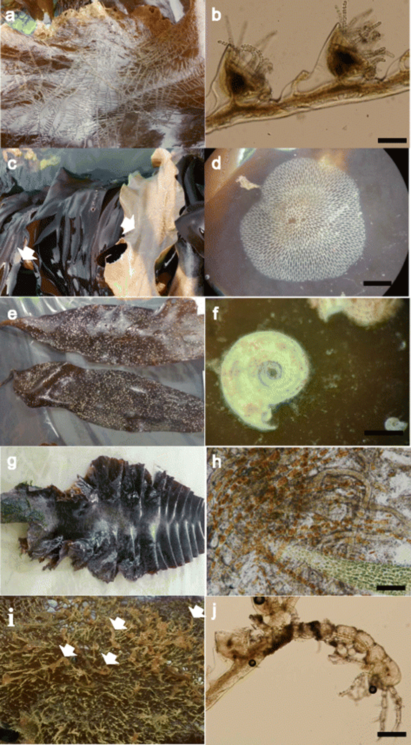
Table 1 shows the results of the detection of epiphytic organisms. Of the 740 kelp samples, 208 had six kinds of epiphytic organisms attached, including hydroid, bryozoan, polychaete, algae, caprellid, and oyster (Fig. 2). Of these, hydroid, bryozoan, polychaete, algae, and caprellid were frequently observed and were detected in the kelp samples with rates of 11.6% (86/740), 6.4% (47/740), 3.4% (25/740), 3.2% (24/740), and 3% (22/740), respectively.

fas-20-0-1-g2
Fig. 2. Photos of epiphytic organisms on cultured kelp. Hydroid (a), bryozoan (c), polychaete (e), algae (g), and caprellid (i) encrusting blades of kelp. Magnification of hydroid (b), bryozoan (d), polychaete (f), algae (h), and caprellid (j). Bar = 100 μm (b, h, and j) and 1000 μm (d, f)
Download Original Figure

Two hundred thirteen samples were examined in the Wando A farm (Table 1). Hydroid, bryozoan, algae, caprellid, and oyster were detected in 7.5% (16/213), 5.6% (12/213), 5.6% (12/213), 2.8% (6/213), and 0.9% (4/213) kelp, respectively, and these organisms were only observed during the period from June to September in 2014. Twenty to 100% were found to be hydroid-positive, 50 to 71.4% were bryozoan-positive, and 14.3 to 100% were algae-positive. Twenty and 71.4% of the caprellid were observed on hydroids, but not on kelp. The oyster (57.1%) was observed in the September 2014 sample. Epiphytic organisms were not observed among the kelp during April to May in 2014 and January to April in 2015. The infestation areas for hydroid, bryozoan, and algae were 10 to 85%, 0.3 to 17%, and 20 to 30%, respectively (Table 2). Two and eight caprellids were observed in the June sample and in the August sample of 2014, respectively.

Table 2. Infestation areas for epiphytic organisms in kelp

Place

Farm

Date

Water temperature (°C)

Mean thalli length (cm)

Mean infection area of epiphytic organisms % (epiphytic area/total area)

Hydroid

Bryozoan

Polychaete

Algae

Caprellida

Oyster

Wando

WDA

Apr 2014

11.5

340 (200–380)

0

0

0

0

0

0

May 2014

15.5

260 (260–270)

0

0

0

0

0

0

Jun 2014

17.4

NT

10 (5–20)

0

0

0

2 (1–5)

0

Jul 2014

21.7

190 (100–240)

85 (70–100)

4 (0.8–7.9)

0

30 (20–50)

0

0

Aug 2014

23

200 (100–240)

81 (60–95)

0.3 (0.2–0.6)

0

20 (10–35)

8 (3–17)

0

Sep 2014

22.1

120 (90–160)

NT

17 (0.2–48.7)

0

0

0

2.35 (0.4–4.3)

Jan 2015

9

41 (20–71)

0

0

0

0

0

0

Jan 2015

8.3

82 (39–122)

0

0

0

0

0

0

Apr 2015

11

236 (200–280)

0

0

0

0

0

0

WDB

May 2014

15.4

220 (100–330)

0

0

0

0

0

0

Jun 2014

21

260 (180–320)

10 (5–25)

0

0

5 (3–6)

0

0

Jul 2014

21.5

160 (130–200)

41.8 (10–80)

0

3 (1–5)

0

2 (1–5)

0

Sep 2014

21.6

130 (60–230)

55 (20–90)

0.13 (0.06–0.5)

0

0

0

0

Feb 2015

9

57 (30–100)

0

0

0

0

0

0

Mar 2015

11

97 (50–180)

0

0

0

0

0

0

Apr 2015

12.5

127 (90–190)

0

0

0

0

0

0

Busan

BS

Apr 2014

11

170 (50–270)

0

0.04 (0.04)

0

0

0

0

May 2014

14.5

230 (130–280)

14.4 (5–30)

7.3 (1.8–14.1)

0

0

0

0

Jun 2014

21

220 (180–300)

34 (10–60)

2.8 (0.03–11.6)

0

34.1 (5–70)

0

0

Jul 2014

20.4

160 (120–180)

37 (20–60)

37.9 (1.1–86)

0

0

3 (1–5)

0

Feb 2015

11.5

76 (26–175)

0

0

0

0

0

0

Feb 2015

12

143 (80–219)

0

0

0

0

0

0

Mar 2015

12.5

182 (110–260)

0

0

0

0

0

0

Apr 2015

13

203 (120–270)

0

0

0

0

0

0

Pohang

PH

Jul 2014

20.5

140 (50–220)

3.3 (3–5)

0.5 (0.5)

9.5 (3–20)

0

0

0

Aug 2014

23.4

110 (80–150)

5.3 (1–10)

1.2 (0.2–3.6)

50 (40–60)

0

0

0

Sep 2014

23.1

70 (30–110)

3 (2–4)

0

90 (80–95)

0

0

0

NT not tested

aMean number of caprellid

Download Excel Table

Two hundred fifty-three samples were examined in the Wando B farm (Table 1). Hydroid, bryozoan, polychaete, algae, and caprellid were detected in 13% (33/253), 2% (5/253), 2% (5/253), 1.6% (4/253), and 0.4% (1/253) kelp, respectively, and these organisms were observed from June to September in 2014. 57.1 to 100% were found to be hydroid-positive, and 33.3, 35.7, and 13.3 to 28.6% were bryozoan-positive, polychaete-positive, and algae-positive, respectively. The caprellid (7.1%) was observed on the hydroids in the July 2014 sample. No epiphytic organisms were observed in the kelp in May 2014 and from February to April 2015. The infestation areas for hydroid gradually increased from 10 to 55% as the water temperature increased, but these were lower than those of the Wando A farm (Table 2). The infestation areas for bryozoan, polychaete, and algae were observed in 0.13% (0.06–0.5%) in the September sample, 3% (1–5%) in the July sample, and 5% (3–6%) in the June sample, 2014, respectively. Two caprellids were observed in the July 2014 sample.

Two hundred thirty-six samples were examined in the Busan farm (Table 1). Hydroid, bryozoan, algae, and caprellid were detected in kelp with an incidence rate of 11% (26/236), 9.7% (23/236), 3.4% (8/236), and 4.2% (10/236), respectively, and these organisms were observed from April to July 2014. One hundred percent of the samples were found to be hydroid-positive from May to July 2014. Ten to 90% and 30 to 71.4% were bryozoan-positive and algae-positive, respectively. No epiphytic organisms were observed among the kelp from February to April 2015. The infestation areas of the hydroid were 14.4 to 37%, which are lower than those of the two farms in Wando (Table 2). In contrast, the infestation areas for bryozoan were of 0.04 to 37.9%, which were the highest in the four farms. 34.1% of the algae were observed in June 2014, and three caprellids were observed in July 2014.

In the Pohang farm, hydroid, bryozoan, and polychaete were detected in kelp with rates of 28.9% (11/38), 18.4% (7/38), and 52.6% (20/38), respectively, from July to September 2014 (Table 1). The infestation areas for hydroid and bryozoan were less than 5.5%, which were the lowest in the four farms (Table 2). In contrast, the infestation areas for polychaete were 9.5 to 90%, which were the highest of the four farms.

Histopathology

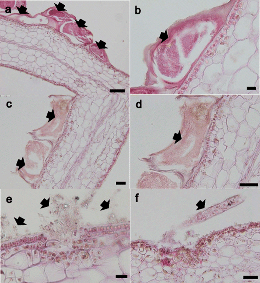
The hydroid, bryozoan, and algae that were attached to the thallus were examined (Fig. 3). The hydroid and bryozoan were attached to the cuticula of the thallus but did not penetrate into the thallus (Fig. 3a–d). In contrast, some of the algae penetrated the epidermis and attached to the cuticula of thallus (Fig. 3e, f).

fas-20-0-1-g3
Fig. 3. Histological section of kelp infected with hydroid (a, b), bryozoan (c, d), and algae (e, f). The arrows indicate the epiphytic organisms. Bar = 20 μm (b, e), 50 μm (c, d, and f), 100 μm (a)
Download Original Figure

Discussion

Korean farms have been cultivating kelp since the 1970s, but to date, no systematic research on their disease has been conducted. In this study, we investigated the presence of epiphytic organisms in four kelp farms in the coastal area of Korea. Of the 740 kelp samples, 208 samples had epiphytic organisms attached, including hydroid, bryozoan, polychaete, algae, caprellid, and oyster. The predominant epiphytic organisms were hydroid (detection rate: 11.6%) followed by bryozoan (6.4%), polychaete (3.4%), algae (3.2%), and caprellid (3%), as observed from May to September. No epiphytic organisms were observed among the kelp from January to April, except for one sample from Busan. These results indicate that at least six kinds of epiphytic organisms were observed in kelp farms in Korea, and their infestation was significantly higher in water with a higher temperature. Moreover, encrusting hydroid and bryozoan were the most predominant form of infestation in kelp farms, even though their infestation rates were different among the kelp farms. Encrusting by hydroid has been reported to be abundant on farmed kelp in Korea at higher water temperatures (Park and Hwang, 2012). The infestation rate from May to July was of about 97%, and it was below 26% from February to April. These results are similar to the results of the present study. Encrusting by bryozoan (Membranipora) has been reported to be abundant on kelp blades in Atlantic Nova Scotia (Canada) and the Gulf of Maine (USA) (Berman et al., 1992; Saunders and Metaxas, 2008; Scheibling and Gagnon, 2009). The calcified Membranipora zooids present a firm mechanical barrier on epidermal tissue. This barrier can affect exchange processes such as mineral nutrient uptake between kelp epidermis and surrounding seawater (Hurd et al., 1994; 2000), or it can interfere with the photophysiology of the host alga (Oswald et al., 1984; Cancino et al., 1987; Muñoz et al., 1991). In this study, even though the effect of the bryozoan on kelp is unknown, Korean kelp farm is suffered from infestation by bryozoan.

It is unclear how the epiphytic organisms attach to the thallus tissue. A histopathogical examination revealed that hydroid and bryozoan were attached on the cuticula of thallus while some algae attached to the cuticula of the thallus or penetrated the epidermis. These results indicate that kelp tissue can be broken by some algae, but not by hydroid and bryozoan.

The cultivation period for kelp in Korea had traditionally spanned from December to July, with a harvest of thalli occurring between June and July for use with edible marine vegetable food. Recently, the harvest season has been shortened from March to May due to the presence of these epiphytic organisms. Moreover, kelp that is severely infected by hydroid and bryozoan is used as abalone feed during the summer, even though their effect on abalone remains. In addition, there are many environmental factors that can affect the prevalence of the epiphytes on kelps. Therefore, further studies are necessary to elucidate which environmental factors are related to the epiphytic organisms’ infestation, to understand how to prevent the epiphytic organisms from infesting kelp farms and to assess their effect when used as abalone feed.

Conclusion

In conclusion, we investigated the presence of epiphytic organisms in four kelp Saccharina japonica farms in the coastal area of Korea from 2014 to 2015. The infestation rate for hydroid, bryozoan, and polychaete was significantly higher in the Wando farm, Busan farm, and Pohang farm, respectively. Epiphytic organisms were generally observed during May to September. The histopathogical examination revealed that hydroid and bryozoan organisms were attached on the cuticula of the thallus. These results indicate that hydroid and bryozoan were the most predominant epiphytic organisms in Korean kelp farms.

Acknowledgements

This study was supported by the National Fisheries Research and Development Institute (R2016069).

Funding

This study was supported by the National Fisheries Research and Development Institute (R2016069).

Availability of data and materials

All datasets in this study are available from the corresponding author on reasonable request.

Authors’ contributions

JOK, WSK, HNJ, and SJC carried out the experiments. JOK and WSK participated to write the manuscript. MJO conceived of the study and participated in its design. JSS and MAP analyzed the data. All authors read and approved the final manuscript.

Competing interests

The authors declare that they have no competing interests.

Consent for publication

Not applicable.

Ethics approval and consent to participate

Not applicable.

References

1.

Berman J, Harris L, Lambert W, Buttrick M, Dufresne M. Recent invasions of the Gulf of Maine: three contrasting ecological histories. Conserv Biol. 1992; 6:435-441

2.

Cancino JM, Muñoz J, Muñoz M, Orellana MC. Effects of the bryozoan Membranipora tuberculata (Bosc.) on the photosynthesis and growth of Gelidium rex Santelices et Abbott. J Exp Mar Biol Ecol. 1987; 113:105-112

3.

FAO (Food and Agriculture Organization of the United Nations). The state of world fisheries and aquaculture. 2014 Rome: Food and Agriculture Department. Food and Agriculture Organization of the United Nations. .

4.

Gong YG, Hwang IG, Ha DS, Hwang MS, Hwang EK, Lee SY, Park EJ. Study on the Saccharina culture technique for abalone feed. 2010; p. 42-49.

5.

Hurd CL, Durante KM, Chia FS, Harrison PJ. Effect of bryozoan colonization on inorganic nitrogen acquisition by the kelps Agarum fimbriatum and Macrocystis integrifolia. Mar Biol. 1994; 121:167-173

6.

Hurd CL, Durante KM, Harrison PJ. Influence of bryozoan colonization on the physiology of the kelp Macrocystis integrifolia (Laminariales, Phaeophyta) from nitrogen-rich and -poor sites in Barkley Sound, British Columbia, Canada. Phycologia. 2000; 39:435-440

7.

Kim HS, Lee KS. Faunal studies on the genus Caprella (Crustacea: Amphipoda, Caprellidae) in Korea. Kor J Zool. 1975; 18:115-126.

8.

KOSIS (Korean statistical information service). 2015. Fishery production survey: statistics by type of fishery and species. Retrieved from http://kosis.kr/eng/statisticsList/statisticsList_01List.jsp?vwcd=MT_ETITLE&parmTabId=M_01_01#SubCont. On March 2016.

9.

Lee CM. A systematic study on Korean Caprellids (Crustacea, Amphipoda) in the east sea. Yongin: Thesis for Master’s degree. Dan Kook University; 1994

10.

Muñoz J, Cancino JM, Molina MX. Effect of encrusting bryozoans on the physiology of their algal substratum. J Mar Biol Assoc UK. 1991; 71:877-882

11.

Oswald RC, Telford N, Seed R, Happey-Wood CM. The effect of encrusting bryozoans on the photosynthetic activity of Fucus serratus L. Estuar Coast Shelf Sci. 1984; 19:697-702

12.

Park JH. Invertebrate fauna of Korea. Athecates. National institute of biological resources ministry of environment; 2011

13.

Park JH. Invertebrate fauna of Korea. Thecates. National institute of biological resources ministry of environment; 2010

14.

Park CS, Hwang EK. Seasonality of epiphytic development of the hydroid Obelia geniculata on cultivated Saccharina japonica (Laminariaceae, Phaeophyta) in Korea. J Appl Phycol. 2012; 24:433-439

15.

Saunders M, Metaxas A. High recruitment of the introduced bryozoan Membranipora membranacea is associated with kelp bed defoliation in Nova Scotia, Canada. Mar Ecol Prog Ser. 2008; 369:139-151

16.

Saunders M, Metaxas A. Effects of temperature, size, and food on the growth of Membranipora membranacea in laboratoy and field studies. Mar Biol. 2009; 156:2267-2276

17.

Scheibling RE, Gagnon P. Temperature-mediated outbreak dynamics of the invasive bryozoan Membranipora membranacea in Nova Scotian kelp beds. Mar Ecol Prog Ser. 2009; 390:1-13

18.

Schwaninger H. Population structure of the widely dispersing marine bryozoan Membranipora membranacea (Cheilostomata): implications for population history, biogeography, and taxonomy. Mar Biol. 1999; 135:411-423

19.

The Korean Society of Systematic Zoology. Seoul: Zoological taxonomy. Jiphyupsa; 2014