Fisheries and Aquatic Sciences
BioMed Central
Research Article

In vitro and in vivo anti-inflammatory activities of a sterol-enriched fraction from freshwater green alga, Spirogyra sp.

Lei Wang1,2, You-Jin Jeon1,2, Jae-Il Kim3,*http://orcid.org/0000-0002-6186-5492
1Department of Marine Life SciencesJeju National University63243JejuSelf-Governing ProvinceRepublic of Korea
2Marine Science InstituteJeju National University63333JejuSelf-Governing ProvinceRepublic of Korea
3Department of Food Science and NutritionPukyong National University48513BusanRepublic of Korea

© The Author(s) 2020. Open AccessThis article is licensed under a Creative Commons Attribution 4.0 International License, which permits use, sharing, adaptation, distribution and reproduction in any medium or format, as long as you give appropriate credit to the original author(s) and the source, provide a link to the Creative Commons licence, and indicate if changes were made. The images or other third party material in this article are included in the article's Creative Commons licence, unless indicated otherwise in a credit line to the material. If material is not included in the article's Creative Commons licence and your intended use is not permitted by statutory regulation or exceeds the permitted use, you will need to obtain permission directly from the copyright holder. To view a copy of this licence, visit http://creativecommons.org/licenses/by/4.0/.

Received: Aug 4, 2020; Accepted: Oct 21, 2020

Published Online: Nov 13, 2020

Abstract

Background

Inflammation plays a crucial role in the pathogenesis of many diseases such as arthritis and atherosclerosis. In the present study, we evaluated anti-inflammatory activity of sterol-rich fraction prepared from Spirogyra sp., a freshwater green alga, in an effort to find bioactive extracts derived from natural sources.

Methods

The sterol content of ethanol extract of Spirogyra sp. (SPE) was enriched by fractionation with hexane (SPEH), resulting 6.7 times higher than SPE. Using this fraction, the in vitro and in vivo anti-inflammatory activities were evaluated in lipopolysaccharides (LPS)-stimulated RAW 264.7 cells and zebrafish.

Results

SPEH effectively and dose-dependently decreased the production of nitric oxide (NO) and prostaglandin E2 (PGE2). SPEH suppressed the production of pro-inflammatory cytokines including interleukin-6 (IL-6), tumor necrosis factor-α (TNF-α), and IL-1β through downregulating nitric oxide synthase (iNOS) and cyclooxygenase-2 (COX-2) expression in LPS-stimulated RAW 264.7 cells without cytotoxicity. The in vivo test results indicated that SPEH significantly and dose-dependently reduced reactive oxygen species (ROS) generation, cell death, and NO production in LPS-stimulated zebrafish.

Conclusions

These results demonstrate that SPEH possesses strong in vitro and in vivo anti-inflammatory activities and has the potential to be used as healthcare or pharmaceutical material for the treatment of inflammatory diseases.

Keywords: Spirogyra sp.; Sterol-enriched fraction; Anti-inflammatory activity; RAW 264.7 cells; Zebrafish

Background

Inflammation is a highly regulated biological process that enables the immune system to efficiently eliminate stimuli and injuries (Masresha et al. 2012). Inflammation can be classified into two subtypes, chronic and acute inflammation, depending on the difference in response time and procedure. Acute inflammation is an initial response of bodies against harmful stimuli. During acute inflammatory response, the plasma and leukocytes are moving from the blood into the injured tissues. There is a shift in the type of mononuclear phagocytic cells in the inflammatory tissues during the course of inflammatory response, from pro-inflammatory to anti-inflammatory or wound healing type.

The inflammatory response is associated with the release of the inflammatory mediators, including nitric oxide (NO), prostaglandins, histamine, and bradykinin from various immune cells such as mononuclear phagocytes and mast cells (González Mosquera et al. 2011; Wijesinghe et al. 2014a). Inflammation is crucial in many diseases such as arthritis, atherosclerosis, cancer, and some deadly diseases (Lee and Weinblatt 2001; Firestein 2006; Klegeris et al. 2007; Wen et al. 2016; Wijesinghe et al. 2013; Wijesinghe et al. 2014b). Therefore, there is increasing attention on finding safe and effective anti-inflammatory agents and elucidating their anti-inflammatory mechanisms.

Algae are considered as a potential bio-resource for the development of functional food, cosmeceutical, and pharmaceutical because they are rich in various bioactive compounds, such as proteins, polysaccharides, phenolic compounds, and sterols (Kim et al. 2016; Sanjeewa et al. 2016; Fernando et al. 2017b). These compounds possess broad-spectrum bioactivities including anti-bacterial, anti-inflammatory, antioxidant, and anti-cancer activities (Jung et al. 2008; Heo et al. 2010; Lee et al. 2012; Lee et al. 2013; Oh et al. 2016; Fernando et al. 2017a).

Spirogyra sp. is freshwater green alga. It has been used as a bio-sorbent to remove heavy metal ions from wastewater (Khalaf 2008). Recently, the pharmacological activities of Spirogyra sp, such as antioxidant, ultraviolet (UV)-protective, and anti-hypertension activities, have been reported (Kang et al. 2015; Lee et al. 2016; Wang et al. 2017). However, the anti-inflammatory activities of Spirogyra sp. have not been evaluated so far. Therefore, in the present study, ethanol extract of Spirogyra sp. (SPE) and the fractions from SPE were prepared, and their anti-inflammatory activities were evaluated in lipopolysaccharides (LPS)-stimulated RAW264.7 cells. The effect of sterol-enriched fraction (SPEH) from SPE on the production of pro-inflammatory mediators was investigated by enzyme-linked immunosorbent assay (ELISA) and western blot analysis. In addition, the in vivo anti-inflammatory effect of SPEH was evaluated using a zebrafish model.

Methods

Chemicals and regents

LPS, acridine orange, 2′,7′-dichlorofluorescin diacetate (DCFH2-DA), dimethyl sulfoxide (DMSO), and diaminofluorescein-FM diacetate (DAF-FM DA) were purchased from Sigma-Aldrich (St. Louis, MO, USA). Fetal bovine serum (FBS), penicillin–streptomycin (P/S), and Dulbecco’s modified Eagle’s medium (DMEM) were purchased from Gibco/BRL (Grand Island, NY, USA). The ELISA kits for measuring prostaglandin E2 (PGE2), interleukin-6 (IL-6), tumor necrosis factor-α (TNF-α), and IL-1β levels were purchased from R&D Systems Inc. (Minneapolis, MN, USA). Antibodies against β-actin, cyclooxygenase-2 (COX-2), and inducible nitric oxide synthase (iNOS) were purchased from Santa Cruz Biotechnology (Santa Cruz, CA, USA). Secondary anti-rabbit and anti-mouse IgG antibodies were purchased from Cell Signaling Technology (Beverly, MA, USA). Other chemicals and regents used in the present study were analytical grade.

Extraction and fractionation

Sampling, processing, extraction, and fractionation were performed following the protocols described in the previous study (Wang et al. 2017). In brief, lyophilized Spirogyra sp. powder was extracted by 70% ethanol; the 70% ethanol extract of Spirogyra sp. (SPE) was fractionated by hexane, chloroform, and ethyl acetate, respectively. Then, the hexane (SPEH), chloroform (SPEC), ethyl acetate (SPEE), and water (SPEW) fractions of SPE were obtained. Samples were concentrated and stored at – 20 °C until used.

Sterol content analysis

The sterol contents of samples were measured by Liebermann Burchard method described by Xiong et al. (2007). In brief, the samples (2 mg) were dissolved in glacial acetic acid (1 mL) and mixed with prepared Liebermann Burchard regent. The mixture was reacted in the dark at the room temperature for 10 min. After reaction, the absorbance of the mixture was recorded at 625 nm using a micro-plate reader (BioTek, Synergy, HT, USA). Cholesterol was used as standard sterol compound to prepare the standard curve.

Cell culture

RAW 264.7 cells (ATCC® TIB-71™) were cultured in DMEM containing FBS (10%) and P/S (1%) under a humidified atmosphere containing 5% CO2 at 37 °C. RAW 264.7 cells were seeded as a density of 1 × 10⁵ cells/mL for experiments.

Determination of NO, pro-inflammatory cytokines, PGE2 production, and cell viability

RAW 264.7 cells were seeded and incubated for 24 h. Cells were treated with different concentrations of samples and incubated for 1 h. After incubation, LPS (1 μg/mL) was introduced to cells. The culture medium was collected and used for measuring NO, PGE2, TNF-α, IL-6, and IL-1β production levels. The NO production levels of LPS-treated RAW264.7 cells were measured using Griess reagent. The PGE2, TNF-α, IL-6, and IL-1β production levels were evaluated using the commercial ELISA kits following the manufacturer’s instructions. The cell viability was measured by MTT assay based on the method described by Wang et al. (2018).

Western blot analysis

The LPS-stimulated RAW 264.7 cells were harvested and lysed. The lysates contain 50 μg of protein were subjected to electrophoresis on SDS–polyacrylamide gels (12%). Then, the proteins were transferred onto nitrocellulose membranes, and the membranes were incubated with the primary antibodies at 4 °C. After 6 h incubation, the membranes were incubated with HRP-conjugated secondary antibody and visualized using ECL reagent (Amersham, Arlington Heights, IL, USA).

Measurement of survival rate and heart-beating rate on LPS-stimulated zebrafish

Approximately 7–9 h post-fertilization (hpf), zebrafish embryos (15 embryos/group) were treated with different concentrations of SPEH for 1 h. Then, LPS (10 μg/mL) was added into the embryo medium. The survival rate was measured at 3 days post-fertilization (dpf) by counting live embryos, and heart-beating rate was measured following the protocol described by Sanjeewa et al. (2018).

Measurement of reactive oxygens species (ROS) generation, cell death, and NO production on LPS-stimulated zebrafish

At 3 dpf, zebrafish were dyed with DCFH2-DA (20 μg/mL), acridine orange (7 μg/mL), and DAF-FM-DA solutions (5 μM) for the detection of ROS generation, cell death, and NO production, respectively. The anesthetized zebrafish were photographed under the microscope equipped with Cool SNAP-Procolor digital camera (Olympus, Japan). The fluorescence intensity of individual zebrafish was quantified using an Image J program.

Statistical analysis

The data were expressed as the mean ± standard error (SE). One-way ANOVA test (using SPSS 11.5 statistical software) was used to compare the mean values of each treatment. The significant differences were established as *p < 0.05, **p < 0.01 as compared to LPS only-treated group and ##p < 0.01 as compared to control group.

Results and discussion

Sterol content of SPE and its fractions

Algae-derived compounds possess various health benefits (Mayer and Hamann 2003; Athukorala and Jeon 2005; Heo et al. 2005; Kotake-Nara et al. 2005). The sterols including cholesterol, β-sitosterol, campesterol, and polyhydroxylated sterols isolated from algae were shown to strong bioactivities, especially anticancer and anti-inflammatory activities (Kazłowska et al. 2013; Elbagory et al. 2015). The freshwater green alga, Spirogyra sp. contains various bioactive compounds. In our previous studies, we have reported antioxidant, UVB photoprotective, and anti-hypertension activities of phenolic compounds isolated from Spirogyra sp. (Kang et al. 2015; Wang et al. 2017). However, the bioactivities of sterols from Spirogyra sp. have not been evaluated yet. In the present study, we have prepared a sterol-enriched fraction (SPEH) from Spirogyra sp. and evaluated its in vitro and in vivo anti-inflammatory activities.

As shown in Fig. 1, the sterol contents of SPE and its fractions were ranged from 0.50% to 9.08%. This result indicates that the sterol content in hexane fraction (SPEH) was enriched with 6.7 times than SPE. Many reports support sterols isolated from algae possess strong anti-inflammation activity (Ku et al. 2013; Sanjeewa et al. 2016; Suh et al. 2018). Our previous study investigated anti-cancer and anti-inflammatory activities of the sterol-rich fraction of Nannochloropsis oculata. The results indicated that the hexane fraction of methanol extract of N. oculata is rich in sterol content and possesses strong anti-inflammatory activity. In this regard, anti-inflammatory activity can be expected from SPEH prepared in this study, in which the content of sterol was 9.08%.

fas-23-0-27-g1
Fig. 1. Sample preparation and anti-inflammatory activity screening. NO generation (b) and cytotoxicity (c) of SPE and its fractions on LPS-stimulated RAW 264.7 cells. NO production was measured by Griess assay and cell viability was measured by MTT assay. The data were expressed as the mean ± SE (n = 3). *p < 0.05, **p < 0.01 as compared to LPS only-treated group and ##p < 0.01 as compared to control group
Download Original Figure

In vitro anti-inflammatory effect of SPEH

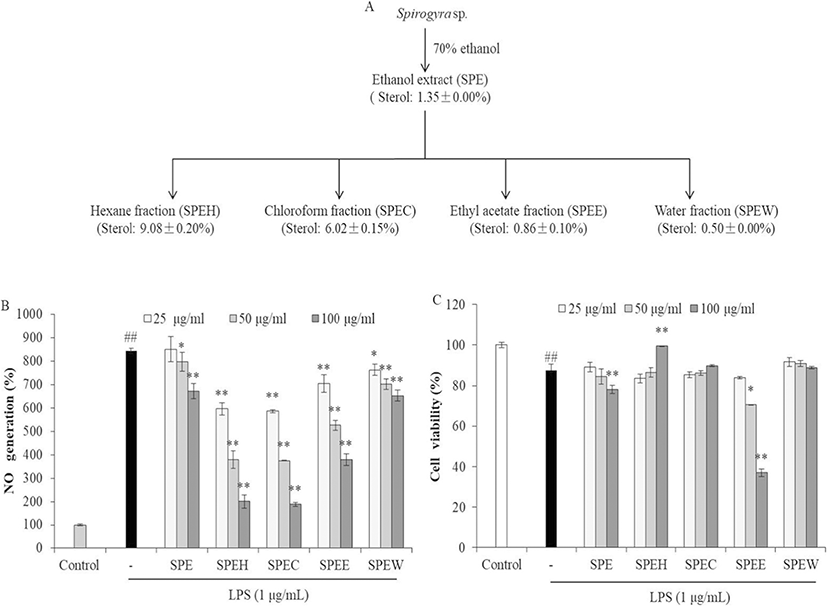
In order to evaluate the anti-inflammatory activity of SPE and its fractions, their inhibitory effects on NO production were measured. As shown in Fig. 1, all samples significantly and dose-dependently decreased NO production (Fig. 1b). However, SPE and SPEE showed remarkable cytotoxicity in RAW264.7 cells. SPEH and SPEC that contain relative higher sterol contents showed stronger NO inhibitory effect in LPS-induced RAW264.7 cells than other samples. Furthermore, SPEH significantly improved the viability of LPS-induced RAW264.7 cells at high concentration. The viability of the cells treated with 100 μg/mL of SPEH was 99.52%, which is close to the cells not treated with LPS (100%) (Fig. 1c). These results indicate that SPEH showed the most potent anti-inflammatory activity among 5 samples. Thus, SPEH was selected as the target sample for the further study.

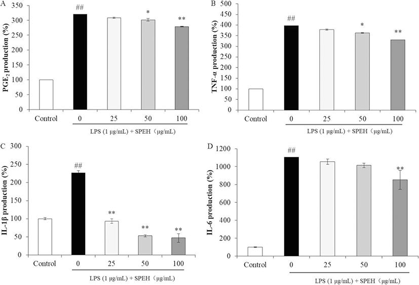
We next examined the inhibitory effects of SPEH on the production of PGE2 and pro-inflammatory cytokines in LPS-stimulated RAW264.7 cells. As Fig. 2a shows, LPS induced PGE2 level in RAW264.7 cells to 319.85% compared to the control group (100%). However, the PGE2 levels of LPS-treated cells were decreased to 307.83%, 301.38%, and 278.90% by 25, 50, and 100 μg/mL of SPEH treatment, respectively. In addition, SPEH inhibited 17.70%, 33.01%, and 65.76% of TNF-α (Fig. 2b), 113.31%, 173.91%, and 179.67% of IL-1β (Fig. 2c), 50.233%, 91.22%, and 252.14% of IL-6 expressions (Fig. 2d) at the concentration of 25, 50, and 100 μg/mL, respectively.

fas-23-0-27-g2
Fig. 2. Inhibitory effect of SPEH on PGE2 (a), TNF-α (b), IL-1β (c), and IL-6 (d) production in LPS-induced RAW 264.7 cells. The PGE2 and pro-inflammatory cytokines levels were determined by ELISA kits according to the manufacturer’s instructions. The data were expressed as the mean ± SE (n = 3). *p < 0.05, **p < 0.01 as compared to LPS only-treated group and ##p < 0.01 as compared to control group
Download Original Figure

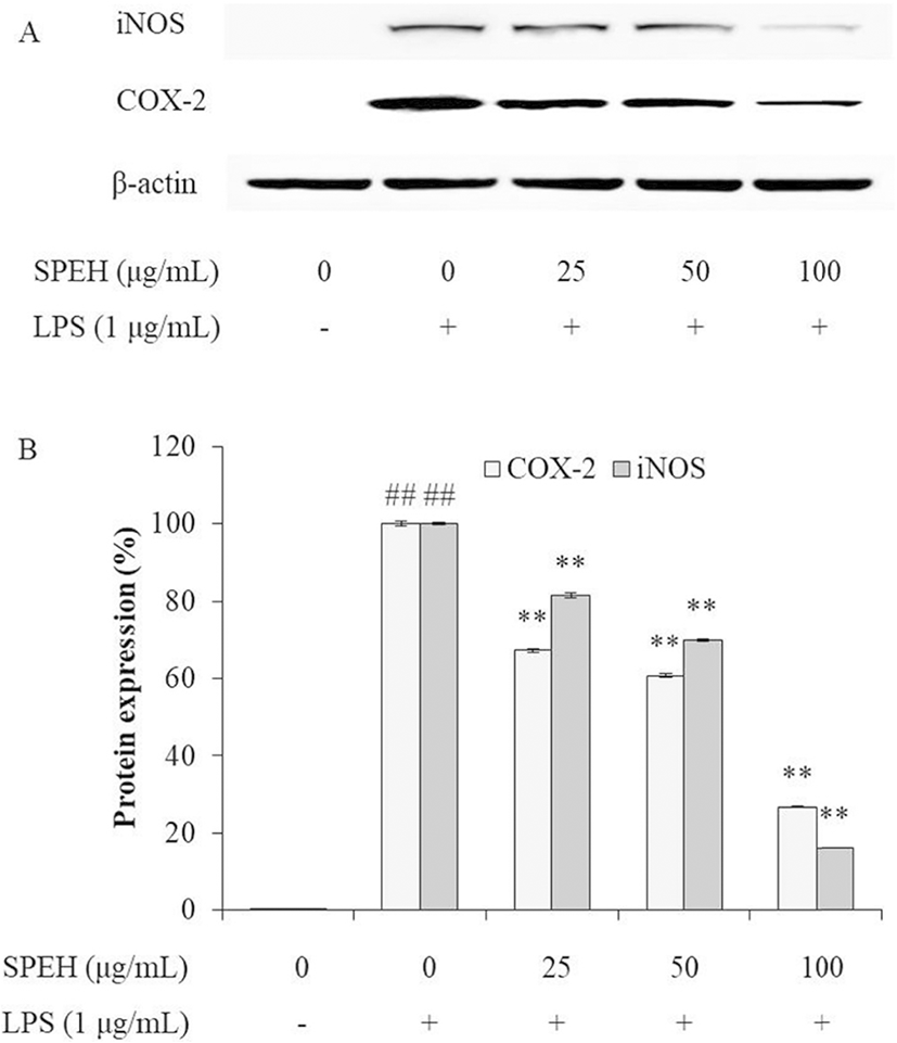
iNOS and COX-2 are two inducible enzymes, which synthesize two key inflammatory mediators, NO and PGE2, respectively (Kim et al. 2005; Wijesinghe et al. 2014a). The levels of iNOS and COX-2 are up-regulated in inflammatory response (Wijesinghe et al. 2014a; Wijesinghe et al. 2014b; Xiong et al. 2014). Therefore, we investigated the effect of SPEH on the protein expression levels of iNOS and COX-2 in LPS-stimulated RAW 264.7 cells. As shown in Fig. 3, LPS significantly stimulated iNOS and COX-2 expressions, but SPEH remarkably and dose-dependently downregulated the expression level of iNOS and COX-2. These results indicated that SPEH possesses strong in vitro anti-inflammatory activity.

fas-23-0-27-g3
Fig. 3. Inhibitory effects of SPEH on iNOS and COX-2 expression in LPS-induced RAW 264.7 cells. a iNOS and COX-2 expression in LPS-induced RAW 264.7 cells. b The relative amount of iNOS and COX-2. Relative amounts of iNOS and COX-2 expressions were compared to β-actin. The data were expressed as the mean ± SE (n = 3). **p < 0.01 as compared to LPS only-treated group and ##p < 0.01 as compared to control group
Download Original Figure

In vivo anti-inflammatory effect of SPEH

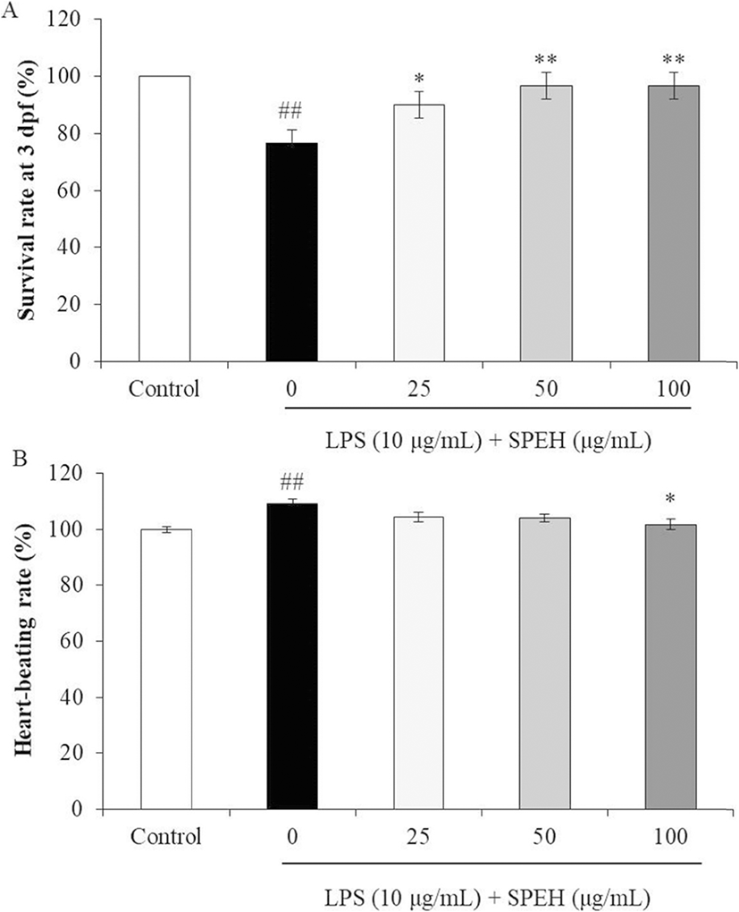
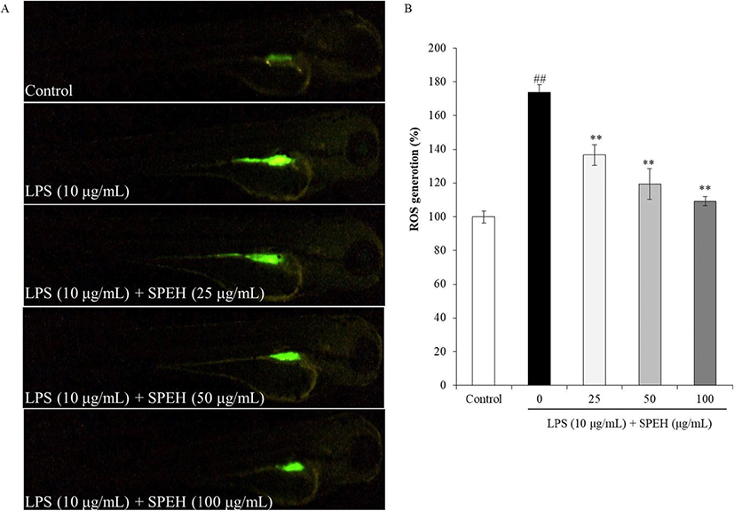
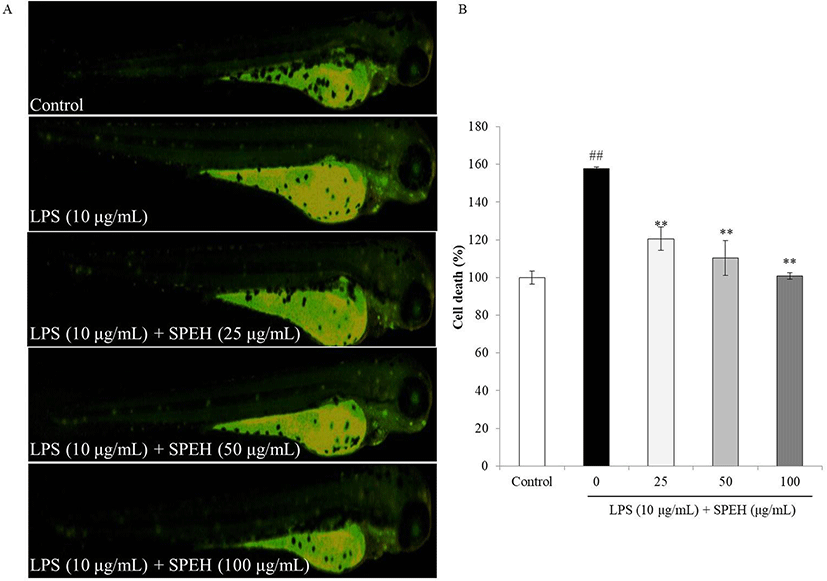
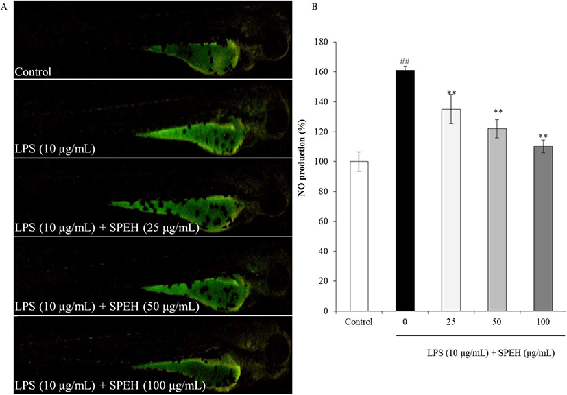
In vivo evaluation for anti-inflammation activity of SPEH was adopted in zebrafish due to its advantages such as similarity to mammals, short life span, and the ability of the female fish to produce a large number of eggs (Kishi et al. 2003; Ko et al. 2011). In the present study, we investigated the survival rate, heart-beating rate, ROS generation, cell death, and NO generation in zebrafish. As shown in Fig. 4a, while the survival rate of zebrafish was significantly reduced by LPS, it was remarkably and dose-dependently increased by SPEH-treatment. In addition, the heart-beating rate of LPS-treated zebrafish was significantly increased (112.93%) compared to non-treated zebrafish (100%) (Fig. 4b). However, it was restored to the normal level in the zebrafish treated with 100 μg/mL of SPEH (101.65%) (Fig. 4b). The ROS generation and cell death levels of SPEH-treated zebrafish were significantly and dose-dependently reduced compared to the zebrafish treated with LPS only (Figs. 5 and 6). Furthermore, SPEH pretreatment reduced 26.04, 39.08, and 50.88% of NO production in LPS-stimulated zebrafish pretreated with 25, 50, and 100 μg/mL, respectively (Fig. 7). These results indicated that SPEH possesses strong in vivo anti-inflammatory activity.

fas-23-0-27-g4
Fig. 4. SPEH improved the survival rate and reduced heart-beating rate in LPS-stimulated zebrafish. a The survival rate. b the heart-beating rate. The data were expressed as the mean ± SE (n = 3). *p < 0.05, and **p < 0.01 as compared to LPS only-treated group and ##p < 0.01 as compared to control group.
Download Original Figure
fas-23-0-27-g5
Fig. 5. The protective effect of SPEH against LPS-induced ROS generation in zebrafish. a Zebrafish under fluorescence microscope. b The levels of ROS. ROS levels were measured using Image J software. The data were expressed as the mean ± SE (n = 3). **p < 0.01 as compared to LPS only-treated group and ##p<0.01 as compared to control group
Download Original Figure
fas-23-0-27-g6
Fig. 6. The protective effect of SPEH against LPS-induced cell death in zebrafish. a Zebrafish under fluorescence microscope. b The levels of cell death. Cell death levels were measured using Image J software. The data were expressed as the mean ± SE (n = 3). **p < 0.01 as compared to LPS only-treated group and ##p < 0.01 as compared to control group
Download Original Figure
fas-23-0-27-g7
Fig. 7. The protective effect of SPEH against LPS-induced NO production in zebrafish. a Zebrafish under fluorescence microscope. b The levels of NO production. NO production levels were measured using ImageJ software. The data were expressed as the mean ± SE (n = 3). **p < 0.01 as compared to LPS only-treated group and ##p < 0.01 as compared to control group
Download Original Figure

Conclusion

In the present study, we prepared a sterol-enriched fraction from Spirogyra sp. (SPEH) and evaluated its in vitro and in vivo anti-inflammatory activities in LPS-stimulated RAW 264.7 cells and zebrafish. The results indicated that SPEH possesses strong anti-inflammatory activity in both in vitro and in vivo models and has the potential to be developed as a healthcare product or a drug to treat inflammatory diseases.

Acknowledgements

Not applicable

Authors’ contributions

LW, YJJ, and JIK conceived and designed the experiments. LW carried out the experiments and wrote the manuscript. YJJ and JIK corrected the manuscript. The authors read and approved the final manuscript.

Funding

This work was supported by a Research Grant of Pukyong National University (2019).

Availability of data and materials

The datasets generated and/or analyzed during the current study are available from the corresponding author on a reasonable request.

Ethics approval and consent to participate

Not applicable

Consent for publication

Not applicable

Competing interests

The authors declare that they have no competing interests.

Publisher’s Note

Springer Nature remains neutral with regard to jurisdictional claims in published maps and institutional affiliations.

References

1.

Athukorala Y, Jeon YJ. Screening for angiotensin 1-converting enzyme inhibitory activity of Ecklonia cava. Prev Nutr Food Sci. 2005; 10:134-139

2.

Elbagory AM, Meyer M, Ali AH, Ameer F, Parker-Nance S, Benito MT, et al. New polyhydroxylated sterols from Palythoa tuberculosa and their apoptotic activity in cancer cells. Steroids. 2015; 101:110-115

3.

Fernando IPS, Kim HS, Sanjeewa KKA, Oh JY, Jeon YJ, Lee WW. Inhibition of inflammatory responses elicited by urban fine dust particles in keratinocytes and macrophages by diphlorethohydroxycarmalol isolated from a brown alga Ishige okamurae. Algae. 2017; 32:261-273

4.

Fernando IPS, Sanjeewa KKA, Le WW, Kim HS, Kim EA, Abeytunga DTU, et al. FTIR characterization and antioxidant activity of water soluble crude polysaccharides of Sri Lankan marine algae. Algae. 2017; 32:75-86

5.

Firestein GS. Inhibiting inflammation in rheumatoid arthritis. N Engl J Med. 2006; 354:80

6.

González Mosquera DM, Ortega YH, Kilonda A, Dehaen W, Pieters L, Apers S. Evaluation of the in vivo anti-inflammatory activity of a flavonoid glycoside from Boldoa purpurascens. Phytochem Lett. 2011; 4:231-234

7.

Heo SJ, Park EJ, Lee KW, Jeon YJ. Antioxidant activities of enzymatic extracts from brown seaweeds. Bioresour Technol. 2005; 96:1613-1623

8.

Heo SJ, Yoon WJ, Kim KN, Ahn GN, Kang SM, Kang DH, et al. Evaluation of anti-inflammatory effect of fucoxanthin isolated from brown algae in lipopolysaccharide-stimulated RAW 264.7 macrophages. Food Chem Toxicol. 2010; 48:2045-2051

9.

Jung WK, Lee DY, Kim JH, Choi IH, Park SG, Seo SK, et al. Anti-inflammatory activity of caffeic acid phenethyl ester (CAPE) extracted from Rhodiola sacra against lipopolysaccharide-induced inflammatory responses in mice. Process Biochem. 2008; 43:783-787

10.

Kang N, Lee JH, Lee WW, Ko JY, Kim EA, Kim JS, et al. Gallic acid isolated from Spirogyra sp. improves cardiovascular disease through a vasorelaxant and antihypertensive effect. Environ Toxicol Pharmacol. 2015; 39:764-772

11.

Kazłowska K, Lin HT, Chang SH, Tsai GJ. In vitro and in vivo anticancer effects of sterol fraction from red algae Porphyra dentata. Evid Based Complement Alternat Med. 2013; 2013:493869

12.

Khalaf MA. Biosorption of reactive dye from textile wastewater by non-viable biomass of Aspergillus niger and Spirogyra sp. Bioresour Technol. 2008; 99:6631-6634

13.

Kim HH, Kim HS, Ko JY, Lee JH, Jeon YJ. A single-step isolation of useful antioxidant compounds from Ishige okamurae by using centrifugal partition chromatography. Fish Aquatic Sci. 2016; 19:22

14.

Kim SF, Huri DA, Snyder SH. Inducible nitric oxide synthase binds, S-nitrosylates, and activates cyclooxygenase-2. Science. 2005; 310:1966-1970

15.

Kishi S, Uchiyama J, Baughman AM, Goto T, Lin MC, Tsai SB. The zebrafish as a vertebrate model of functional aging and very gradual senescence. Exp Gerontol. 2003; 38:777-786

16.

Klegeris A, McGeer EG, McGeer PL. Therapeutic approaches to inflammation in neurodegenerative disease. Curr Opin Neurol. 2007; 20:351-357

17.

Ko SC, Cha SH, Heo SJ, Lee SH, Kang SM, Jeon YJ. Protective effect of Ecklonia cava on UVB-induced oxidative stress: in vitro and in vivo zebrafish model. J Appl Phycol. 2011; 23:697-708

18.

Kotake-Nara E, Asai A, Nagao A. Neoxanthin and fucoxanthin induce apoptosis in PC-3 human prostate cancer cells. Cancer Lett. 2005; 220:75-84

19.

Ku CS, Pham TX, Park Y, Kim B, Shin MS, Kang I, et al. Edible blue-green algae reduce the production of pro-inflammatory cytokines by inhibiting NF-κB pathway in macrophages and splenocytes. Biochim Biophs Acta. 2013; 1830:2981-2988

20.

Lee DM, Weinblatt ME. Rheumatoid arthritis. Lancet. 2001; 358:903-911

21.

Lee JC, Hou MF, Huang HW, Chang FR, Yeh CC, Tang JY, et al. Marine algal natural products with anti-oxidative, anti-inflammatory, and anti-cancer properties. Cancer Cell Int. 2013; 13:55

22.

Lee JH, Han JW, Ko JY, Lee WW, Ahn G, Kim CY, et al. Protective effect of a freshwater alga, Spirogyra sp., against lipid peroxidation in vivo zebrafish and purification of antioxidative compounds using preparative centrifugal partition chromatography. J Appl Phycol. 2016; 28:181-189

23.

Lee SH, Ko CI, Ahn G, You SG, Kim JS, Hue MS, et al. Molecular characteristics and anti-inflammatory activity of the fucoidan extracted from Ecklonia cava. Carbohydr Polym. 2012; 89:599-606

24.

Masresha B, Makonnen E, Debella A. In vivo anti-inflammatory activities of Ocimum suave in mice. J Ethnopharmacol. 2012; 142:201-205

25.

Mayer AMS, Hamann MT. Marine pharmacology in 2000: marine compounds with antibacterial, anticoagulant, antifungal, anti-inflammatory, antimalarial, antiplatelet, antituberculosis, and antiviral activities; affecting the cardiovascular, immune, and nervous systems and other miscellaneous mechanisms of action. Mar Biotechnol (NY). 2003; 6:37-52

26.

Oh JY, Fernando IPS, Jeon YJ. Potential applications of radioprotective phytochemicals from marine algae. Algae. 2016; 31:403-414

27.

Sanjeewa KKA, Fernando IPS, Kim SY, Kim HS, Ahn G, Jee Y, et al. In vitro and in vivo anti-inflammatory activities of high molecular weight sulfated polysaccharide; containing fucose separated from Sargassum horneri: short communication. Int J Biol Macromol. 2018; 107:803-807

28.

Sanjeewa KKA, Fernando IPS, Samarakoon KW, Lakmal HHC, Kim EA, Kwon ON, et al. Anti-inflammatory and anti-cancer activities of sterol rich fraction of cultured marine microalga Nannochloropsis oculata. Algae. 2016; 31:277-287

29.

Suh SS, Hong JM, Kim EJ, Jung SW, Kim SM, Kim JE, et al. Anti-inflammation and anti-cancer activity of ethanol extract of antarctic freshwater microalga, Micractinium sp. Int J Med Sci. 2018; 15:929-936

30.

Wang L, Oh JY, Kim HS, Lee WW, Cui Y, Lee HG, et al. Protective effect of polysaccharides from Celluclast-assisted extract of Hizikia fusiforme against hydrogen peroxide-induced oxidative stress in vitro in Vero cells and in vivo in zebrafish. Int J Biol Macromol. 2018; 112:483-489

31.

Wang L, Ryu BM, Kim WS, Kim GH, Jeon YJ. Protective effect of gallic acid derivatives from the freshwater green alga Spirogyra sp. against ultraviolet B-induced apoptosis through reactive oxygen species clearance in human keratinocytes and zebrafish. Algae. 2017; 32:379-388

32.

Wen ZS, Xiang XW, Jin HX, Guo XY, Liu LJ, Huang YN, et al. Composition and anti-inflammatory effect of polysaccharides from Sargassum horneri in RAW264.7 macrophages. Int J Biol Macromol. 2016; 88:403-413

33.

Wijesinghe WAJP, Ahn G, Lee WW, Kang MC, Kim EA, Jeon YJ. Anti-inflammatory activity of phlorotannin-rich fermented Ecklonia cava processing by-product extract in lipopolysaccharide-stimulated RAW 264.7 macrophages. J Appl Phycol. 2013; 25:1207-1213

34.

Wijesinghe WAJP, Kang MC, Lee HS, Kamada T, Vairappan CS, Jeon YJ. 5β-Hydroxypalisadin B isolated from red alga Laurencia snackeyi attenuates inflammatory response in lipopolysaccharide-stimulated RAW 264.7 macrophages. Algae. 2014; 29:331-341

35.

Wijesinghe WAJP, Kim EA, Kang MC, Lee WW, Lee HS, Vairappan CS, et al. Assessment of anti-inflammatory effect of 5β-hydroxypalisadin B isolated from red seaweed Laurencia snackeyi in zebrafish embryo in vivo model. Environ Toxicol Pharmacol. 2014; 37:110-117

36.

Xiong H, Cheng Y, Zhang X, Zhang X. Effects of taraxasterol on iNOS and COX-2 expression in LPS-induced RAW 264.7 macrophages. J Ethnopharmacol. 2014; 155:753-757

37.

Xiong Q, Wilson WK, Pang J. The Liebermann–Burchard reaction: sulfonation, desaturation, and rearrangment of cholesterol in acid. Lipids. 2007; 42:87-96