

Introduction
Parasitism is a common phenomenon in marine ecosystems, where nearly all biota, especially fish, are susceptible to parasitic infections. The parasite life cycle involves organisms ranging from zooplankton and fish to marine mammals, serving as both paratenic and definitive hosts. Some parasite species also have zoonotic potential, infecting humans as accidental hosts and causing diarrhea, nausea, vomiting, and abdominal pain (Bao et al., 2019). Helminth endoparasites have evolved specialised adaptations for parasitism, including reproductive strategies and transmission within intermediate, paratenic, and definitive hosts to complete their life cycle. Host diet and food preferences play a crucial role in the transmission of endoparasites and the establishment of community structure.
Anisakis sp. is one of the most common helminth endoparasites that widely spreads with a heteroxenous life cycle comprising four larval stages (L1–L4) (Klimpel & Palm, 2011). Anisakis sp. has been found infecting small pelagic fish commodities, such as Indian mackerel (Rastrelliger spp.), along the southern coast of East Java (Setyobudi et al., 2019). Another study on the phylum Acanthocephala, Rhadinoryncus sp., reported infections in commercial fish species in Japan, including tuna, mackerel, and salmon (Yamaguti, 1963). Adult larvae are found in the digestive system, including the intestine, pyloric caeca, and the external surfaces of some internal organs, of the infected Serrasentis sagittifer (Abdel-Ghaffar et al., 2014). Interactions among fish as hosts within the life cycle of helminth endoparasites closely relate to ecological conditions, host distribution, and the host’s adaptive capabilities, which influence the geographical distribution of the parasites (Marcogliese, 2008).
Small pelagic fish are among the main and highly abundant catches in the coastal waters of The Special Region of Yogyakarata. Rastrelliger sp., Sardinella sp., and Selar sp. are small pelagic fish caught year-round and play a crucial role in the marine ecosystem and the regional economy. These fish have been reported to be susceptible to endoparasitic helminth infections, which can affect fish health and potentially reduce marketability. Several studies have examined helminth endoparasite infections in marine fish commodities in Indonesia. However, a significant gap remains in understanding the diversity and distribution of helminth species across fish species.
Studies on parasite community infections are essential for understanding parasite epidemiology and supporting the development of parasites as biological indicators, which are crucial for fisheries resource management and conservation (Dobson et al., 2008). Therefore, this study aims to identify the helminth endoparasite species infecting small pelagic fish, determine their prevalence and infection intensity, and assess the host diet to understand the structure of the parasite community. This research is essential for improving the understanding of epidemiology, life cycles, biogeography, and phylogeny of helminth endoparasites, particularly those from the southern coast of The Special Region of Yogyakarta.
Materials and Methods
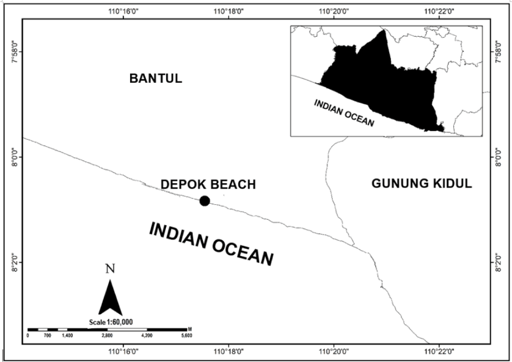
Helminth endoparasite larvae were collected from 100 small pelagic fish caught from Depok Beach in the southern coast of Yogyakarta (Fig. 1), during November 2024–March 2025. Each fish sampled was measured for body weight and total length and identified according to Collette & Nauen (1983). Helminth endoparasite larvae collected were examined through dissection and examination of the abdominal cavity, digestive tract, liver, and gonads. The collected larvae were washed repeatedly in physiological saline solution, then stored in 70% ethanol for morphological and molecular identification. The parasite population infection was calculated according to the method described by Bush et al. (1997).
A total of two helminth larvae (an Anisakis sp. and a Rhadinorhynchus sp.) that have been preserved in 70% ethanol were examined using a scanning electron microscope (SEM) the next day in Laboratorium Penelitian dan Pengujian Terpadu (LPPT), Universitas Gadjah Mada. Prior to observation, the specimens were removed from ethanol, air-dried at room temperature, and placed on SEM stubs. No additional fixation, dehydration series, or conductive coating was applied. SEM observations focused on the anterior and posterior regions to observe diagnostic morphological features. Morphological observations for Anisakis sp. refer to Van Hien et al. (2021), and for Rhadinorhynchus sp., to Kita et al. (2024) for reference on obtaining SEM images.
Genomic DNA was extracted from helminth larvae using the Genomic DNA Mini Kit Tissue (Favorgen, Ping Tung, Taiwan) according to the manufacturer’s instructions. Furthermore, for Anisakis larvae, the internal transcribed spacer (ITS) region (ITS1-5.8S-ITS2) of rDNA was amplified using primers A (5'-GTC GAA TTC GTA GGT GAA CCT GCG GAA GGA TCA-3') and B (5'-GCC GGA TCC GAA TCC TGG TTA GTT TCT TTT CCT-3') (D’Amelio et al., 2000). DNA amplification was then carried out using PCR with the composition reaction of 0.5 µL primer A and B, 6 µL Kapa Mix (Kapa Biosystem, Wilmington, MA, USA), 5 µL nuclease free water, and 0.5 µL of template DNA under conditions of pre-denaturation at 94°C for 10 minutes, 35 cycles of denaturation for 40 seconds, annealing at 55°C for 40 seconds, extension at 72°C for 90 seconds, final extension at 72°C for 7 minutes. For Rhadinorhynchus larvae, the 18S region of rDNA was amplified using primers 18S rRNA SSU ribosomal nucleotides using Univ-Prst-F (5’-ACG-GCT-ACC-ACA-TCC-AAG-GAA-3) and Univ-Prst-R (5’-TGT-ACA-AAG-GGC-AGG-GAC-GTA-3’) (Murwantoko & Hayati, 2022) under conditions of pre-denaturation at 94°C for 10 minutes, 35 cycles of denaturation for 40 seconds, annealing at 60°C for 40 seconds, extension at 72°C for 90 seconds, final extension at 72°C for 7 minutes. The PCR products were analyzed by electrophoresis in a 1.5% agarose gel supplemented with 0.75 μL of Fluorescent DNA stain (1st base). Electrophoresis was run for 25 minutes, and the result was photographed using a UV Transilluminator (Pacific Image Electronic, New Taipei City, Taiwan). The PCR product was then sequenced using a DNA sequencing (Sanger) service company (Singapore’s 1st Base Laboratory through PT. Genetika Science Indonesia). Furthermore, the alignment nucleotide sequence was analyzed using the basic local alignment search tool (BLAST) on the NCBI website (ncbi.nlm.nih.gov), and the phylogenetic tree was constructed using MEGA XI (Tamura et al., 2021). Phylogenetic relationships were analyzed using the maximum-likelihood tree with bootstrap test phylogeny (1,000 replicates) and the Kimura 2-parameter model.
The digestive tract of the examined fish was measured. The volume of each food type was measured using a modified Sedgwick Rafter. Each food type was observed using quantitative methods (numerical and volumetric), then examined under a microscope at magnifications of 4× to 10×, and identified using a plankton identification book (Shirota, 1966). Data analysis, including relative gut length, frequency of occurrence, and index of preponderance.
- Relative gut length (RGL) (Al-Hussaini, 1949).
Interpretation:
RGL > 3: herbivorous
RGL = 1–3: omnivorous
RGL < 1: carnivorous
- Frequency of occurrence (FO; Effendie, 1979).
(Ni, total number of one type of organism; I, total number of gut-filled)
- Index of preponderance (IP; Biswas, 1993)
(Vi, percentage of food volume; Oi, percentage of occurrence)
The index of preponderance is divided into three groups: main food (IP > 25%), complementary food (5% ≤ IP ≤ 25%), and additional food (IP < 5%).
Results
One hundred specimens of small pelagic fish from three species were examined. Samples comprise Selar crumenophthalmus (N = 38), Sardinella gibbosa (N = 30), and Rastrelliger faughni (N = 32), with sizes varying as shown in Table 1.
Nematodes (Anisakis sp.) and acanthocephalans (Rhadinorhynchus sp.) were found infecting small pelagic fish. Anisakis sp. was detected in all examined species, whereas Rhadinorhynchus sp. was observed only in the intestine of R. faughni, with a prevalence of 15.63% and a mean intensity of 2.0 larvae per fish (Table 2). Anisakis sp. exhibited the highest prevalence across all host species, with the highest value in R. faughni ([P] = 28.13%), followed by S. crumenophthalmus (P = 21.05%), and the lowest in S. gibbosa (16.67%). These findings indicate that Anisakis sp. is more widely distributed among the studied fish species, infecting both the visceral cavity and gonads, whereas Rhadinorhynchus sp. appears to be host-specific and restricted to a single species, with comparatively lower prevalence.
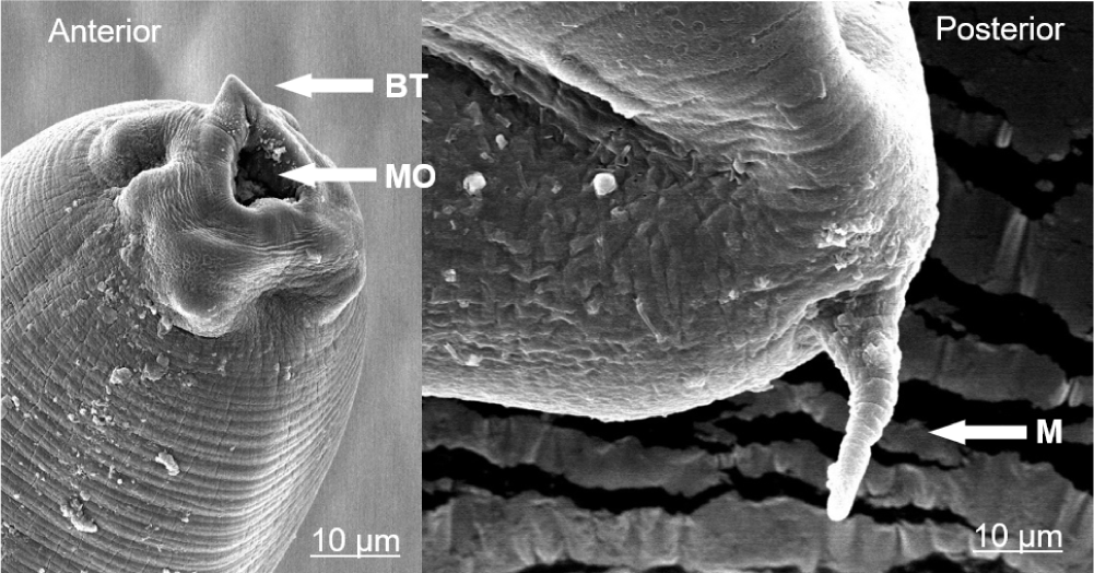
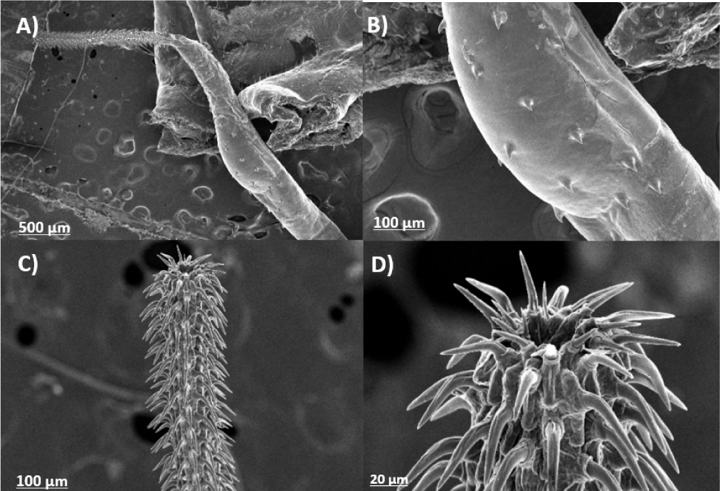
Morphological identification indicates that Anisakis infects all examined fish species, which were Anisakis Type I, characterized by a translucent white, cylindrical body. SEM confirmed the presence of a cylindrical mucron, narrow, and long in the posterior end. The anterior end has a triangular mouth, and the anterior end has a boring tooth as shown in Fig. 2. Rhadinorhynchus specimens collected from R. faughni showed typical acanthocephalan morphology, including a cylindrical, unsegmented body with a retractable proboscis armed with longitudinal rows of recurved hooks (Fig. 3). The proboscis is long, cylindrical, and slightly widening anteriorly, with 35 longitudinal rows of 11–14 hooks for each. The proboscis contained 35 longitudinal rows of hooks, with 11–14 hooks per row.
The direct sequencing of the ITS region of rDNA from six Anisakis specimens collected from each fish species resulted in diverse sizes of nucleotide sequences ranging from 915 to 922 bp. Molecular identification shows that the Anisakis larvae infecting R. faughni, S. crumenophthalmus, and S. gibbosa were Anisakis typica. Rhadinorhynchus specimens from R. faughni were also identified by direct sequencing of the 18S RNA, and the result shown the closest similarity with Rhadinorhynchus dorsoventrosiponosus. The relationships among the helminth endoparasites isolated in this study and the sequence data published in GenBank shown in Fig. 4.
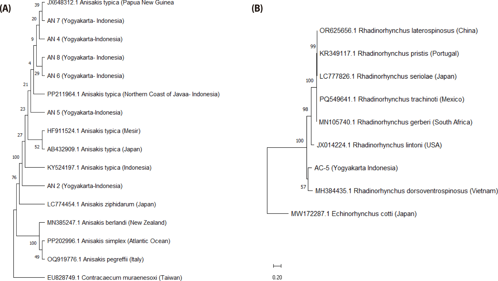
Anisakis phylogeny revealed that samples from Yogyakarta, Indonesia (coded AN-2, AN-4, AN-5, AN-6, AN-7, and AN-8) clustered within the same clade as A. typica reference sequences from multiple geographic regions. Other Anisakis species formed a distinct subclade, with Contracaecum muraenesoxi from Taiwan serving as the outgroup. Rhadinorhynchus phylogeny demonstrated that specimens infecting R. faughni consist of two clades, one of which consists of Rhadinorhynchus spp. and Echynorhynchuscotti as an outgroup.
The dietary analyses of R. faughni, S. crumenophthalmus, and S. gibbosa, from the southern coast of The Special Region of Yogyakarata, are all carnivorous, characterized by relatively short gut lengths (Table 3). The gut length of three fish species ranges from 3.30 to 29.8 cm, with relative gut lengths ranging from 0.17 to 0.97 cm.
Stomach content analysis revealed distinct and overlapping prey preferences among the three small pelagic fish species. R. faughni and S. gibbosa primarily consumed a diverse range of zooplankton with total percentage (50.85% and 90.38%), including Calanus spp., Euterpina spp., Temora spp., Ceratium spp., Corycaeus spp., Sagitta spp., Cyclops spp., Porcellidium spp., phytoplankton including Surirella spp., Chaetoceros spp., and Peridium spp., and eggs. S. crumenophthalmus exhibited a broader diet, incorporating larger prey items such as fish, crustaceans, and molluscs. Shrimp and fish (in whole form, as pieces of fish flesh, and as fish bones) were common dietary components for all three species, indicating some overlap in their trophic niches. The food composition from each fish species examined is shown in Fig. 5.
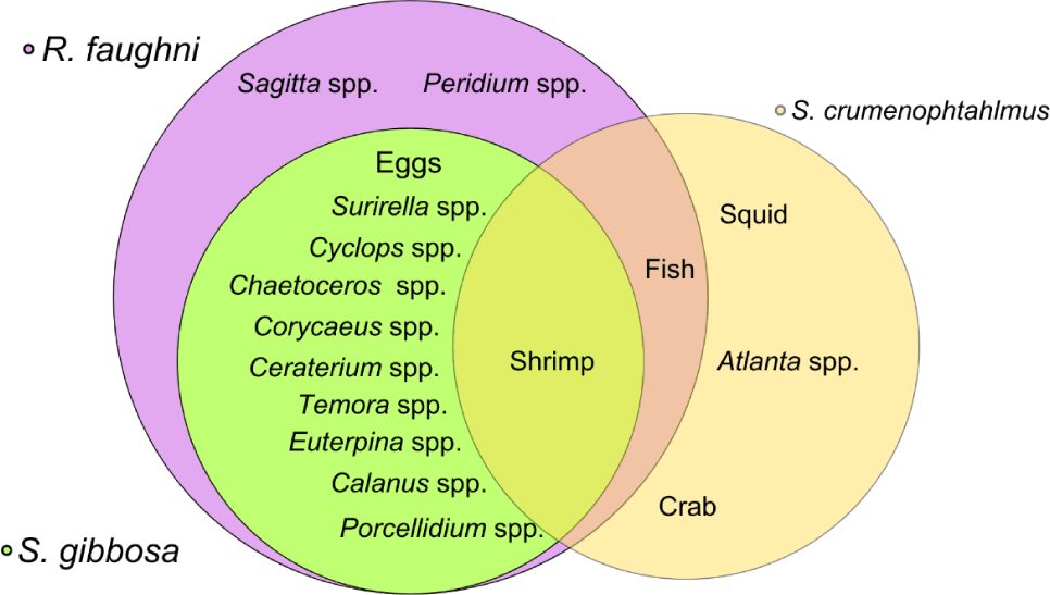
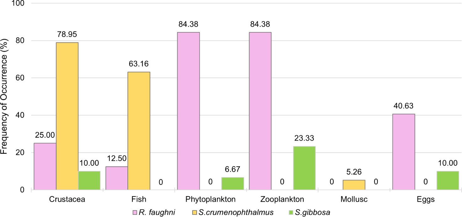
Phytoplankton and zooplankton are the two most common types of food found in the digestive tracts of R. faughni and S. gibbosa. Crustaceans were the only food type found in all three fish species, with frequencies of 10.00% in S. gibbosa, 25.00% in R. faughni, and the highest at 78.95% in S. crumenophthalmus. Shrimp were found in the stomachs of all three species, either whole or in pieces, whereas crabs were only found in S. crumenophthalmus (Fig. 6).
Zooplankton was the primary food for R. faughni and S. gibbosa, with IP values of 56.86% and 97.50%, respectively. In contrast, crustaceans were the primary food source for S. crumenophthalmus (83.55%), with fish as the secondary source (16.04%). Crustaceans and zooplankton were the dominant food types for the three small pelagic fish species in the southern coast of The Special Region of Yogyakarata, as shown in Table 4.
Discussion
R. faughni appears to be the most common and diverse parasitised host among the three species studied, primarily infecting the visceral cavity, intestine, and gonad, and harbouring both parasite taxa. These findings differ from those of Setyobudi et al. (2019), who reported a lower prevalence (P = 17%) but a higher intensity ([MI] = 6.8 larvae/fish) of Anisakis infection in Rastrelliger spp. from the southern coast of East Java. According to Mattiucci et al. (2008), such differences in prevalence among the same host species may indicate that host populations originate from different stocks. Parasite distribution and infection levels are influenced by the presence and distribution of hosts within the parasite life cycle, migration patterns, and the abundance of paratenic or definitive hosts (Kuhn et al., 2016).
The distribution of Anisakis infection sites in small pelagic fish was predominantly in the body cavity. Rohde (1984) noted that Anisakis most frequently accumulates in the digestive tract because it uses its boring tooth to migrate to visceral organs, even penetrating muscle and flesh. In contrast, Rhadinorhynchus larvae were only detected in the intestines of R. faughni. The detection of Rhadinorhynchus in R. faughni represents the first report of this parasite in this commodity in Indonesia. According to Amin (2020), adults of Rhadinorhynchus have been reported to infect the intestines of various fish families, including Carangidae, Salmonidae, Scombridae, Terapontidae, Tetraodontidae, and Trichiuridae.
Morphological observations of Anisakis larvae revealed elongated cylindrical bodies with a translucent appearance. Further, SEM observations show that Anisakis larvae have a triangular mouth equipped with a boring tooth. At the posterior end, a prominent terminal cylindrical mucron was observed. According to Marcogliese (2008), the Anisakis species found in the three small pelagic fish species correspond morphologically to Anisakis Type I. The protruding posterior mucron and the mouth opening with a boring tooth and a triangular thick lip are characteristic of A. typica, distinguishing it from other species (Marcogliese, 2008).
The nucleotide sequence from the rDNA ITS region was compared with the NCBI database, and all Anisakis samples identified shared more than 99.75% sequence similarity with A. typica from various geographic locations. The six Anisakis samples exhibited high similarity to A. typica originating from Indonesia, Egypt, Japan, China, Thailand, and Papua New Guinea. Previous studies have confirmed that A. typica is the most common Anisakis species infecting fish in Indonesian waters (Anshary et al., 2014; Palm et al., 2017; Setyobudi et al., 2019). The population of A. typica has been genetically detected across a wide geographic range, spanning from 35° to 40°N and from 35°S to the tropics in both temperate and tropical waters (Mattiucci et al., 2008). According to Mattiucci et al. (2008), A. typica reaches adulthood by infecting various dolphin species within the families Delphinidae and Phocoenidae in warm and tropical waters, serving as definitive hosts in its life cycle.
Anisakis samples from the southern coast of The Special Region of Yogyakarata (coded AN-2 to AN-8) were in the same subclade as A. typica from various global locations. A. typica with codes AN-4, AN-6, AN-7, and AN-8 have the closest phylogenetic relationships to A. typica from Papua New Guinea, and AN-5 has the closest phylogeny with A. typica from the northern coast of Java. The European Food Safety Authority (EFSA Panel on Biological Hazards, 2010) notes that the worldwide distribution of Anisakis means no marine area is free of this parasite, due to its migratory nature and heteroxenous life cycle, which involves multiple hosts.
The Acanthocephalan larvae infecting R. faughni have elongated, cylindrical bodies, are whitish, and larger than Anisakis larvae. SEM of the acanthocephalan samples revealed a relatively long proboscis with hooks distributed on both the ventral and dorsal surfaces. This morphology corresponds to Rhadinorhynchus spp., where hooks are distributed from the proboscis to the abdomen, separated by aspinose areas or connected by lateral hooks (Amin et al., 2019). The proboscis of Rhadinorhynchus sp. in this study consisted of 35 longitudinal rows of hooks, with 11–17 hooks per row. This number exceeds previous descriptions by Amin et al. (2019) and Kita et al. (2024), reported that Rhadinorhynchus generally has 8–26 longitudinal rows with 17–48 hooks per row. The ventral side has 28 hooks, while the dorsal’s hooks could not be observed. According to Amin et al. (2011), the number of ventral hooks in Rhadinorhynchus ranges from 11 to 43, and dorsal hooks range from 7 to 27. The anterior part of this larvae covered with hooks, serves as an anchoring organ that attaches to the intestinal wall and prevents dislodgement during digestion (Amin et al., 2019).
Species-level identification was performed by DNA amplification of Rhadinorhynchus targeting the 18S SSU rRNA region, yielding a DNA fragment of 1,115 base pairs. BLAST analysis against the NCBI database showed that the identified Rhadinorhynchus sample shared 84.69% sequence similarity with Rhadinorhynchus dorsoventrospinosus, with a query coverage of 69%. This reference sequence was collected from Decapterus kurroides in Vietnamese waters. Molecular diversity and phylogeny of the genus Rhadinorhynchus remain poorly characterized, and information on the infection and distribution of this parasite genus is limited. According to Kita et al. (2024), taxonomic studies on this genus are underdeveloped due to frequent misidentifications and a lack of traceability of fish capture locations.
Phylogenetic analysis placed specimen AC-5 from Yogyakarta close to R. dorsoventrospinosus, supported by a bootstrap value of 57%, confirming that AC-5 is R. dorsoventrospinosus. The phylogenetic tree revealed branching comprising the genus Rhadinorhynchus from several geographic locations within a single clade. Echinorhynchus cotti served as the outgroup, belonging to a different genus within the same family, Rhadinorhynchidae. Kita et al. (2024) reported that R. dorsoventrospinosus clusters are genetically close to R. seriolae and R. hiansi. The molecular diversity and phylogeny of the genus Rhadinorhynchus remain insufficiently understood. Only ten Rhadinorhynchus species have been identified, with six others still uncharacterized (Kita et al., 2024).
Diet analysis is important for understanding the distribution and transmission of endoparasites in marine fish. Food webs play both functional and structural roles in the life cycles of parasites, which are closely linked to ecological conditions. The three fish species examined had gut lengths ranging from 3.30 to 29.80 cm, indicating they are classified as carnivorous. R. faughni exhibited the most diverse diet, with nearly all food groups represented, except for molluscs. Zooplankton was the most abundant food type found in the stomachs of R. faughni, with Euterpina spp. being the most abundant and common species, at 24.15%. According to Hakimelahi et al. (2020), Rastrelliger spp. in the Persian Gulf tend to consume zooplankton, phytoplankton, algae, and molluscs. The presence of plankton in the diet of Rastrelliger spp. across various waters indicates that this fish is carnivorous and tends towards planktivory. Similarly, S. gibbosa exhibited a plankton-feeding preference, with zooplankton of the genus Temora comprising 30.99% of the stomach contents. In contrast, S. crumenophthalmus predominantly consumed crustaceans, mainly shrimp (65.35%), and was the only species that consumed molluscs, including squid (3.96%) and Atlanta spp. (1.98%).
Crustaceans were the only food type found in all three fish species, with frequencies of 10.00% in S. gibbosa, 25.00% in R. faughni, and the highest at 78.95% in S. crumenophthalmus. Shrimp were found in the stomachs of all three species, either whole or in pieces, whereas crabs were only found in S. crumenophthalmus. Roux & Conand (2000) reported that adult S. crumenophthalmus in La Réunion primarily feed on fish and crustaceans such as shrimp, whereas smaller Selar species showed higher frequencies of shrimp, crab, and euphausiids than other food types. Zooplankton were the primary food source for R. faughni and S. gibbosa, with IP values of 56.86% and 97.50%. In comparison, crustaceans were the primary food source for S. crumenophthalmus (83.55%), with fish serving as the secondary food source (16.04%). According to Syahputra et al. (2016), differences in fish diet proportions can be caused by variations in food availability, fish behavior, and environmental factors that influence food resources.
Parasite identification also showed that Anisakis infected all three species, and R. faughni additionally harboured Acanthocephala from the genus Rhadinorhynchus. According to Marcogliese (1995), nematode parasites and some genera of Acanthocephala infect zooplankton during their life cycles. Oshima et al. (1969) noted that zooplankton such as euphausiids (krill) serve as crucial intermediate hosts in the life cycle of Anisakis. Larvae moult to the L2 stage in the water and then enter small zooplankton and crustaceans as the first intermediate hosts before infecting fish. Subsequently, larvae enter larger marine crustaceans, cephalopods, and fish, where they encyst as third-stage larvae (L3) before maturing into fourth-stage larvae (L4) and reproducing in definitive hosts, namely cetaceans.
In contrast, the life cycle of Rhadinorhynchus remains poorly understood. Grano-Maldonado et al. (2024) indicated that definitive hosts are fish, where the parasite reproduces in the digestive tract. Eggs are released into the water with the host’s faeces, ingested by small crustacean intermediate hosts, and hatch into acanthor larvae. These develop into second-stage larvae (acanthella) and cystacanths (L3 or juvenile). In Acanthocephala, these stages can occur within the same host until the infected intermediate host is consumed by a fish, at which point the cystacanths mature into adults and reproduce (Buchmann & Karami, 2024).
Parasite transmission can occur among various zooplankton taxa without involving specific species. The zooplankton identified in the diet of R. faughni and S. gibbosa from the southern coast of The Special Region of Yogyakarata included copepod genera Calanus, Euterpina, Temora, Corycaeus, Cyclops, and Porcellidium, as well as Sagitta from the class Sagittoidea. Marcogliese (1995) reported that copepods and malacostracans are known as intermediate hosts of Anisakis spp. The diverse diet of R. faughni, compared with that of the other two fish species, is associated with infections by A. typica and Rhadinorhynchus sp. Costa et al. (2000) found Rhadinorhynchus infections in Trachurus picturatus, a species that typically consumes small crustaceans. Kennedy (2006) noted that malacostracans, including mysids and euphausiids, act as intermediate hosts for Rhadinorhynchus.
The presence of Anisakis and Rhadinorhynchus infections in pelagic fish commodities from the southern coast of The Special Region of Yogyakarata indicates that this area is endemic for these parasites. Mackenzie & Abaunza (1998) defined endemic areas as geographic regions with conditions conducive to parasite transmission. This principle underlies the use of parasites, especially Anisakis, as biological markers to distinguish fish stocks and understand migration patterns (MacKenzie, 2002). According to Marcogliese (2008), environmental conditions, host distribution, and parasite adaptability are the main factors influencing the geographic distribution of Anisakis spp. Host distribution is the most critical factor, as the presence of intermediate, paratenic, and definitive hosts determines the occurrence of parasites in a given area, the absence of hosts prevents the completion of the parasite life cycle (Mattiucci et al., 2008). Furthermore, infection of helminth endoparasites in fish is influenced by the fish’s diet, as individual dietary preferences affect exposure to transmitted parasites (Bolnick et al., 2003).
Conclusion
Helminth endoparasites infecting three small pelagic fish species on the southern coast of The Special Region of Yogyakarta were identified as A. typica and Rhadinorhynchus sp., with only Rhadinorhynchus sp. found in R. faughni. Molecular analysis indicated that Rhadinorhynchus showed the highest similarity to R. dorsoventrospinosus, although the confidence was relatively low, and further identification is required. The highest prevalence of Anisakis sp. was recorded in R. faughni (28.13%), followed by S. crumenophthalmus (21.05%) and S. gibbosa (16.67%), while Rhadinorhynchus prevalence in R. faughni was 15.63%. The average infection intensity of Anisakis was 1.67 larvae/fish in R. faughni, 1.25 larvae/fish in S. crumenophthalmus, and the highest in S. gibbosa (3.20 larvae/fish). In contrast, Rhadinorhynchus in R. faughni reached 2.00 larvae/fish. Anisakis was mainly found in the abdominal cavity and gonads, while Rhadinorhynchus was restricted to the intestine, and no helminth endoparasites were detected in the muscle tissue.
Research on helminth endoparasite infections should be expanded to include a wider range of fish species across different geographical regions and conducted over defined temporal scales. Periodic monitoring of infection distribution, particularly of Anisakis spp., is essential for understanding parasite population dynamics, fish migration patterns, and the application of these parasites as biological tags. Host feeding habits are also necessary to accurately determine intermediate hosts involved in the parasite life cycle. Furthermore, molecular identification of Rhadinorhynchus spp. should incorporate additional genetic markers to improve taxonomic resolution and ensure accurate species-level identification.


