

Introduction
Bone is a dynamic, highly vascularized tissue that undergoes continuous remodeling in response to mechanical and metabolic stimuli (Laubach et al., 2022). It comprises a calcified matrix-approximately 65% inorganic material, 25% organic material, and 10% water-together with cellular components (osteoblasts, osteoclasts, and osteocytes) and regulatory proteins such as osteocalcin, osteopontin, and osteoprotegerin (Kim et al., 2022; Laubach et al., 2022). Although bone has a remarkable regenerative capacity and can restore its original structure and function after minor injuries (e.g., fractures and small defects), critical-sized bone defects caused by trauma, tumor resection, infection, congenital malformation, or degenerative diseases exceed its intrinsic healing capacity and therefore require clinical intervention (Wang et al., 2023; Wang et al., 2021b; Yang et al., 2023).
Traditional treatment strategies, including autografts, allografts, and xenografts, remain the standard of care; however, they present significant limitations, such as donor-site morbidity, immune rejection, infection risk, and limited tissue availability (Wang et al., 2022b; Zhang et al., 2023). Against this backdrop, bone tissue engineering has emerged as a promising alternative that seeks to develop bioinspired scaffolds capable of recapitulating the structural, mechanical, and biological features of native bone (Li et al., 2024; Li et al., 2022; Shen et al., 2023). An ideal scaffold should not only promote the expression of biomarkers (e.g., osteocalcin, osteopontin, and osteoprotegerin) but also support mineralization, which is a critical for restoring the architecture and function of damaged bone (Wang et al., 2021b; Zhou et al., 2021).
Recent advances in fabrication techniques, including electrospinning and three-dimensional (3D) printing, have enabled the development of biomimetic scaffolds that more closely resemble the extracellular matrix and support both osteogenic differentiation and angiogenesis (Banimohamad-Shotorbani et al., 2021; Zhang et al., 2023; Zhou et al., 2021). In addition, incorporating functional agents such as bioceramics, anti-inflammatory drugs, and anti-bacterial drugs has further improved scaffold performance by enhancing osteogenic marker expression, promoting mineralization, and even supporting neuroregeneration (Heo et al., 2019; Kim et al., 2024; Shen et al., 2023).
Biomaterials play a central role in contemporary regenerative strategies, serving as scaffolds, drug carriers, and bioactive matrices that interact with biological systems to promote tissue repair and regeneration (Hosseini et al., 2023; Kim et al., 2023; Kurdi & M-Ridha, 2023). These materials are engineered to replace or support damaged tissues, facilitate healing, and ultimately restore function (Oleksy et al., 2023;Zheng et al., 2023). In particular, biomaterials for bone regeneration are designed to mimic the structural and mechanical characteristics of natural bone, thereby supporting the repair of defects arising from trauma, disease, or congenital conditions (Szwed-Georgiou et al., 2023; Xing et al., 2023). Their capacity to support cellular activities, promote osteogenesis, and undergo controlled degradation makes them essential for developing effective bone-regenerative therapies.
Polycaprolactone (PCL) is a synthetic, biodegradable polymer that has attracted considerable attention in tissue engineering, especially for bone regeneration (Javkhlan et al., 2024; Oh et al., 2021). This semi-crystalline aliphatic polyester has been approved by the United States Food and Drug Administration for various biomedical applications owing to its favorable biocompatibility, mechanical integrity, and slow degradation rate (Kim et al., 2023; Wang et al., 2022a). However, PCL is intrinsically hydrophobic, which limits cell adhesion and bioactivity due to poor surface wettability. These drawbacks can be mitigated by incorporating bioactive components such as calcium carbonate (CaCO3, CC), tricalcium phosphate, or marine-derived collagen to enhance osteoconductivity and cell-material interactions (Erdemli et al., 2024; Ferreira et al., 2012).
CC is a biofunctional material widely investigated for bone regeneration because of its biocompatibility, biodegradability, and osteoconductivity (Erdemli et al., 2024; Reyna-Urrutia et al., 2023; Wang et al., 2021a). By providing an inorganic phase analogous to that of native bone, CC can improve osteoblast attachment and activity. In addition, compared with hydroxyapatite, CC often exhibits more favorable biodegradability that can better match the pace of physiological remodeling (Jiang et al., 2023; Liu & Wang, 2023; Xu et al., 2021). Accordingly, CC-containing scaffolds have shown promising outcomes in supporting bone formation and regeneration (Erdemli et al., 2024; Liu & Wang, 2023; Xu et al., 2021). Nevertheless, commercially available CC is typically produced via synthesis and resource-intensive processing, raising concerns regarding cost and environmental sustainability.
To address these limitations, we explored an alternative CC source derived from the marine organism Crossaster papposus japonicus, an abundant yet ecological underutilized species. CC extracted from C. papposus japonicus (CCJ) can be obtained as a by-product of existing from existing marine resource-utilization activities, thereby avoiding the need for dedicated harvesting. Although collection and freeze-drying still incur processing costs, the absence of raw-material procurement costs and the relative simplicity of the extraction procedure may reduce overall expenses compared to conventional chemically synthesized CC (c-CC). Moreover, CCJ may achieve comparable or improved biological performance at lower loading levels than c-CC, which could further enhance cost efficiency. Importantly, valorizing a marine by-product aligns with sustainability goals and supports the development of cost-effective, high-performance scaffolds for clinical translation.
Therefore, the primary objective of this study was to extract and comprehensively characterize CCJ and to fabricate a PCL-based composite scaffold using 3D printing. We further aimed to systematically evaluate the physicochemical properties and biological performance of the resulting PCL/CCJ scaffold and to assess its feasibility as a cost-effective, bioactive, and sustainable biomaterial platform for guided bone regeneration.
Materials and Methods
C. papposus japonicus was collected from the deep-sea floor (depth range: 300–1,000 m) near Wangdol-cho, located in the southwestern East Sea, during a trawl survey conducted by the National Institute of Fisheries Science in June 2017. Ethanol, sodium hypochlorite, sodium hydroxide, and sodium phosphate were purchased from Ducksan Chemical (Ansan, Korea). α-Minimum Eagle’s Medium (α-MEM), fetal bovine serum (FBS), and penicillin/streptomycin/amphotericin were obtained from Gibco (NY, USA). 1-StepTM p-NPP, 3-(4,5-dimethylthiazol-2-yl)-2,5-diphenyltetrazolium bromide (MTT), dimethyl sulfoxide (DMSO), PCL, Alizarin Red S, ascorbic acid, β-glycerophosphate, and cetylpyridinium chloride were purchased from Sigma-Aldrich (St. Louis, MO, USA). All other chemicals and reagents were commercially available.
CCJ was extracted following a previously reported method with minor modifications (Kurdi & M-Ridha, 2023). Briefly, C. papposus japonicus was washed with tap water, rinsed with distilled water, and frozen at –80°C. The frozen samples were lyophilized and homogenized into a fine powder. A total of 100 g of powder was treated with 2.5 L of 70% ethanol to remove soluble impurities. The mixture was filtered through filter paper (GE Healthcare, Chicago, IL, USA), and the residue was collected. The residue was then treated with sodium hypochlorite and stirred for 12 h to eliminate remaining organic components, followed by filtration. Subsequently, the residue was treated with sodium phosphate for 2 h to induce CCJ precipitation. The suspension was centrifuged (25,000×g, 3 mins), and the resulting pellet was washed thoroughly with distilled water. The final CCJ powder was dried in a drying oven to remove residual moisture and stored at –80°C until use.
To identify the functional groups present in CCJ and c-CC, Fourier transform infrared (FT-IR) spectra were acquired using an FT-IR spectrometer (JASCO, Tokyo, Japan). Samples were prepared using the conventional KBr pellet method. Spectra were collected over 650–4,000 cm-1 at a resolution of 4 cm-1 with 30 scans.
X-ray diffraction (XRD) analysis was performed using an XRD diffractometer (X’Pert3 Powder, Malvern Panalytical, Almelo, Netherlands) with Cu-Kα radiation. Diffraction patterns were recorded over a 2θ range of 5° to 90° at a scanning rate of 2.4°/min to assess the crystalline structure and phase purity of CCJ and c-CC.
Cytotoxicity and effect on in vitro differentiation of the CC from C. papposus japonicus (CCJ)
MC3T3-E1 pre-osteoblasts were obtained from the American Type Culture Collection (Rockville, MD, USA) and cultured in α-MEM supplemented with 10% FBS and penicillin/streptomycin/amphotericin. Cells were maintained at 37°C in a humidified incubator with 5% CO2 and subcultured every 3 days.
For cytotoxicity testing, MC3T3-E1 pre-osteoblasts were seeded in 24-well plates at a density of 1 × 10⁵ cells/well. After 24 h, the cells were treated with CCJ at various concentrations (0–200 μg/mL in α-MEM) and incubated for 2 and 4 days. Cell viability was determined using an MTT assay. Briefly, 100 μL of MTT solution (1 mg/mL in PBS) was added to each well and incubated for 2 h at 37°C. Formazan crystals were dissolved in DMSO, and absorbance was measured at 540 nm using a microplate reader.
For alkaline phosphatase (ALP) activity, 100 μL of 1-StepTM p-NPP reagent was added to each well after 7 days of incubation and allowed to react for 30 min at RT. The reaction was terminated by adding 2 N NaOH, and absorbance was measured at 405 nm.
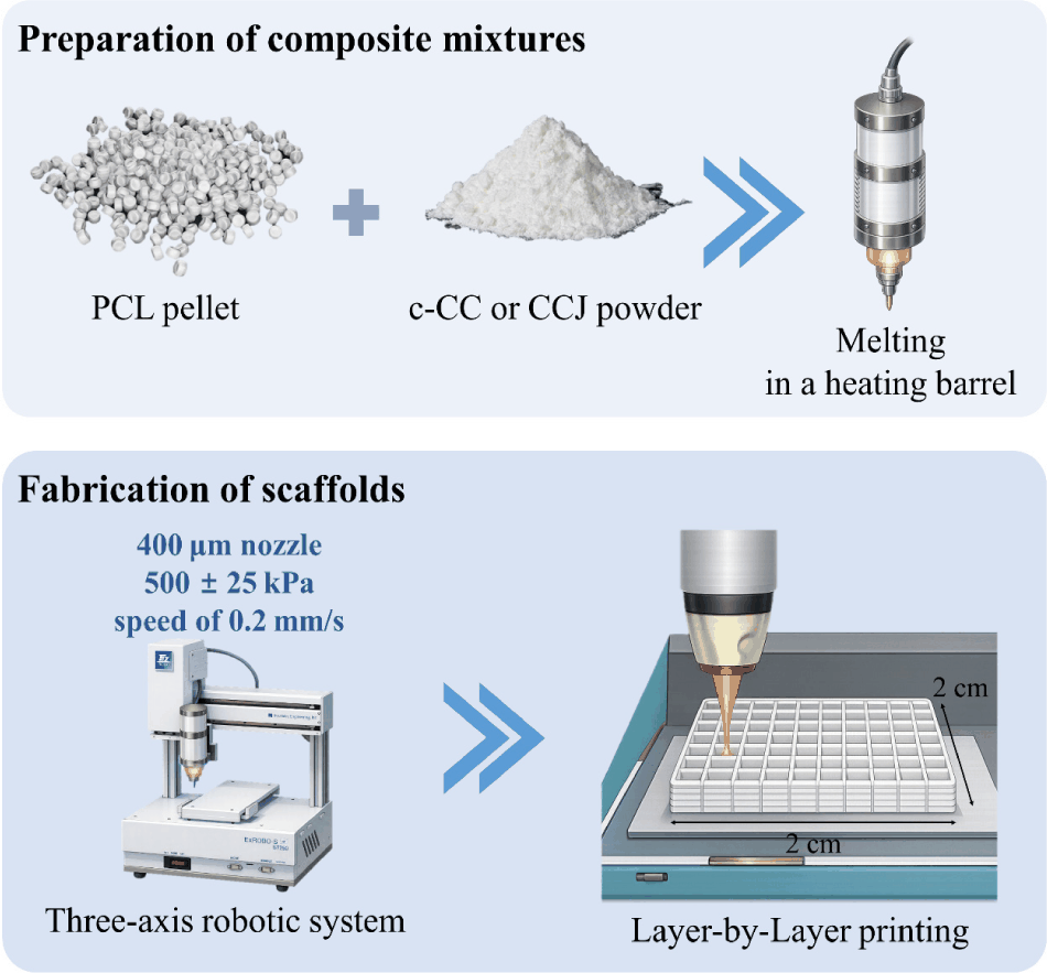
A three-axis robotic plotting system (EZ-ROBO-5GX ST2520, Iwashita Engineering, Kitakyushu, Japan), equipped with a dispenser was used to fabricate scaffolds as previously described (Heo et al, 2019; Kim et al., 2022). As shown in Fig. 1, PCL pellets were premixed with either c-CC or CCJ powder (1% w/w each), and the blended mixtures were melted in a heating barrel to obtain composite melts. The melts were extruded through a 400 μm heated nozzle at a constant pressure of 500 ± 25 kPa and a dispensing speed of 0.2 mm/s. The molten materials were deposited in a layer-by-layer manner to produce 3D scaffolds with uniform pore architecture. Each scaffold was fabricated at a size of 2 × 2 cm and subsequently trimmed for further experiments.
Calcium deposition was assessed by Alizarin Red S staining. Prior to cell seeding, scaffolds were sterilized with 70% ethanol and exposed to UV light for 30 min. MC3T3-E1 cells were seeded onto the scaffolds (0.5× 0.5 mm) at a density of 1 × 10⁵ cells/scaffold. Cells were cultured in osteogenic differentiation medium supplemented with 50 μg/mL ascorbic acid and 10 mM β-glycerophosphate, with medium changes every 2 days. After 7 and 14 days, cells were fixed with cold 70% ethanol and stained with 40 mM Alizarin Red S (pH 4.2) for 30 min at RT. Samples were then washed three times to remove excess stain. For quantification, the stained calcium deposits were eluted with cetylpyridinium chloride, and absorbance was measured at 562 nm using a microplate reader.
All data are presented as the mean ± standard deviation from at least three independent experiments. Statistical significance was evaluated using one-way analysis of variance (ANOVA) followed by Duncan’s multiple range test. Analyses were performed using SPSS Statistics (version 12.0; Chicago, IL, USA). Statistical significance was defined as *p < 0.05 and **p < 0.10.
Results and Discussion
CCJ was successfully extracted using sodium hypochlorite pre-treatment followed by sodium phosphate treatment, and its characteristics were evaluated by FT-IR and XRD. The FT-IR spectra of c-CC and CCJ are shown in Fig. 2A. Both samples exhibited characteristic carbonate bands, including the ν3 asymmetric stretching vibration of CO32- at 1,400 cm-1 and the ν4 in-plane bending vibration at 725 cm-1 (Naknonhan et al., 2025). In addition, peaks at 1,080 cm-1 and 874 cm-1 corresponded to carbonate vibrational modes (Rodriguez-Blanco et al., 2011). In c-CC, a broad band in the range of 2,850–3,050 cm-1 was observed and attributed to O-H stretching vibrations.
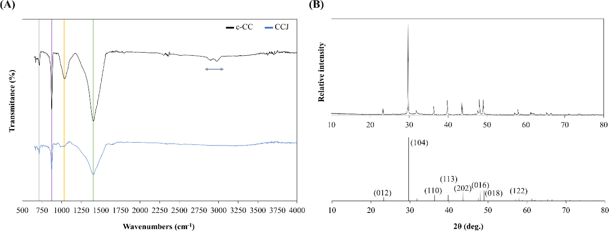
The XRD pattern of the CCJ (Fig. 2B) indicated a highly crystalline phase, with diffraction peaks consistent with calcite calcium carbonate (JCPDS card No. 01-072-1650) (Liu et al., 2018). No additional peaks beyond those assigned to CaCO3 were detected, suggesting that CCJ exhibits a phase purity comparable to that of commercial c-CC. Collectively, these results confirm that CC was successfully extracted from CCJ and that its chemical features are comparable to those of c-CC, supporting CCJ as a potential biomaterial for bone regeneration applications.
The cytotoxicity of CCJ was assessed using the MTT assay. MC3T3-E1 pre-osteoblasts were treated with CCJ (0–200 μg/mL) for 2 and 4 days. As shown in Fig. 3A, CCJ did not reduced cell viability at any tested concentration, indicating no detectable cytotoxicity under these conditions.
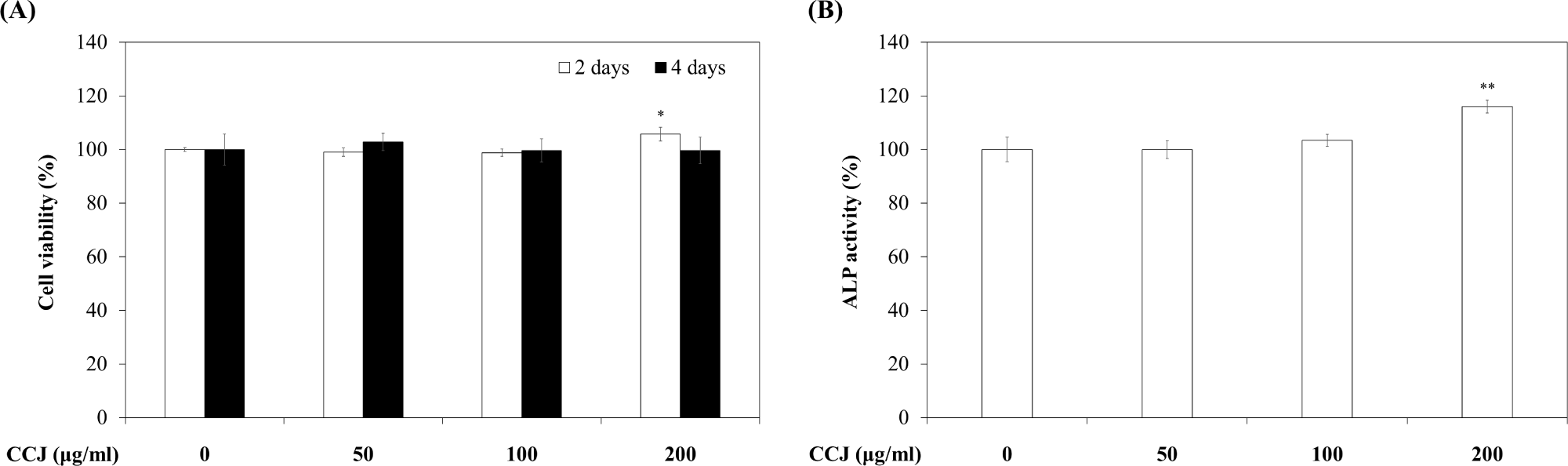
Induction of osteogenic differentiation is an important criterion for bone-regeneration biomaterials. ALP, an early osteogenic marker, contribute to phosphate metabolism by catalyzing the hydrolysis of phosphomonoester bonds (Heo et al, 2019; Xu et al, 2021). Accordingly, the effect of CCJ on ALP activity was evaluated in MC3T3-E1 cells. As shown in Fig. 3B, treatment with 200 μg/mL CCJ for 7 days increased ALP activity relative to the untreated control, showing a statistical trend (p < 0.1). This elevation suggests the early initiation of osteoblast differentiation, potentially associated with increased calcium ion availability from CCJ (Amamoto et al., 2022; Assefa et al., 2022; Jeon et al., 2021).
Overall, these findings suggest that CCJ can support osteogenic responses, potentially by providing bioavailable calcium and enhancing cell-matrix interactions (Erdemli et al., 2024; Wang et al., 2021a). Moreover, the osteogenic performance of PCL/CCJ scaffolds may reflect synergistic effects between the osteoconductive CCJ phase and the mechanical stability provided by the PCL matrix, which together create a favorable microenvironment for osteogenic commitment (Kim et al., 2024; Oh et al., 2021).
Mineralization is a key function of osteoblasts during bone regeneration (Ponzetti & Rucci, 2021). To evaluate mineralization in vitro, calcium deposition by MC3T3-E1 cells cultured on the fabricated scaffolds was assessed at 7 and 14 days using Alizarin Red S staining. As shown in Fig. 4, PCL/CCJ scaffolds enhanced calcium deposition compared with PCL and PCL/c-CC scaffolds, with deposition increasing over time. The PCL/CCJ group exhibited the highest mineralization at both time points.
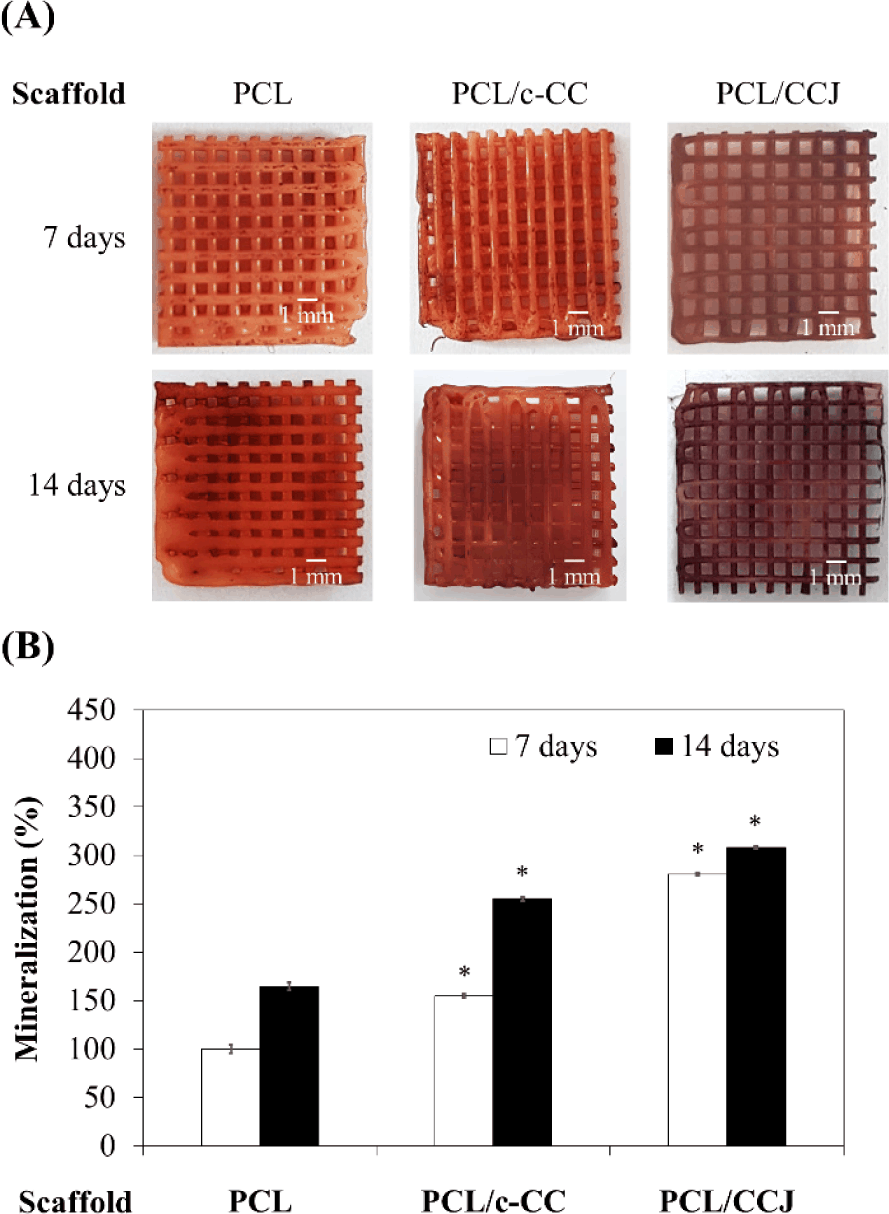
These results indicate that PCL/CCJ scaffolds promotes extracellular matrix mineralization, likely due to the combined contributions of osteoconductive CCJ and the structural integrity provided by the PCL framework. The greater effect observed for CCJ relative to c-CC may be related to differences in bioceramic particle characteristics, such as particle size and morphology, which can influence osteogenic performance (Hayashi et al., 2020). Therefore, future studies will examine the impacts effects of particle size and related physicochemical parameters on osteogenic outcomes in greater detail. Collectively, these findings demonstrate the strong in vitro bone-regenerative potential of the PCL/CCJ scaffold.
Conclusion
In this study, CC was successfully extracted from C. papposus japonicus and incorporated into a morphologically uniform PCL/CCJ scaffold. In vitro evaluation using MC3T3-E1 pre-osteoblasts showed that CCJ increased ALP activity without detectable cytotoxicity. Moreover, the PCL/CCJ scaffold significantly enhanced extracellular matrix mineralization in vitro. Collectively, these findings highlight the potential of marine-derived CCJ as a bioactive filler and support the PCL/CCJ scaffolds as a promising candidates for bone regeneration.


