

Introduction
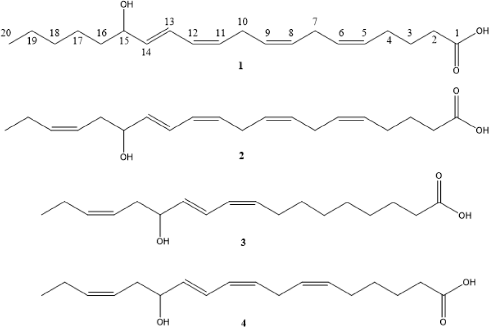
Eisenia bicyclis is an edible alga commonly used in South Korea and is distributed throughout the temperate regions of South Korea and Japan (Kang et al., 2001; Kim et al., 2012; Jung et al., 2013). It is a perennial brown alga belonging to the family Lessoniaceae, which has been previously reported to generate various metabolites such as phlorotannins (eckol, dieckol, phlorofucofuroeckol A, bieckols, and phloroeckols), sterols (fucosterol, saringosterol, and 24-ketocholesterol), and carotenoid (fucoxanthin) (Kim et al., 2011; Kim et al., 2013; Kurata et al., 1990; Moon et al., 2011; Negara et al., 2021; Okeke et al., 2021). Phlorotannins (polyphenolic compounds) are commonly polymerized by 1,3,5-trihydrobenzene (phloroglucinol) and are classified into six subclasses (phloroethols, fucols, eckols, carmalols, fucophloroehols, and fuhalols) (Chen et al., 2021; Duan et al., 2023; Go et al., 2024). They are key bioactive substances originated from E. bicyclis (Sugiura et al., 2021). These metabolites have been shown to possess potent bio-activities including anti-inflammatory, anti-oxidant, anti-viral, anti-diabetic, anti-cancer, and anti-microbial activities (Khan et al., 2022; Lee & Jeon, 2013; Lemesheva et al., 2023; Maheswari & Babu, 2022; Pradhan & Ki, 2023; Shin et al., 2014). In this current paper, we focus on the isolation, structural determination, quantification, and anti-inflammatory activity of four oxylipin derivatives (1–4, Fig. 1) obtained from 70% ethanol extract of E. bicyclis.
Materials and Methods
Nuclear magnetic resonance (NMR) spectra were recorded using a VNMRS 500 MHz Fourier-transform nuclear magnetic resonance (FT-NMR) spectrometer (Varian, Palo Alto, CA, USA). The high-resolution quadrupole time-of-flight mass spectrometry (HR-Q-TOF-MS) spectra were obtained using a SCIEX X500R quadrupole time-of-flight (Q-TOF) liquid chromatography–tandem mass spectrometry (LC-MS/MS) spectrometer (SCIEX, Marlborough, MA, USA). High-performance liquid chromatography (HPLC) was performed using Waters ACQUITY Arc HPLC system (Waters, Milford, MA, USA). All chemical solvents were obtained by HPLC grades (J.T.baker, Radnor, PA, USA).
The brown alga, E. bicyclis was obtained from Jindo-gun, Jeallanam-do, Republic of Korea. A voucher specimen has been deposited at the National Marine Biodiversity Institute of Korea (MABIK).
The brown alga E. bicyclis was washed and freeze-dried, subsequently, the dried E. bicyclis (400 g) was ground and extracted three times with 70% ethanol (2 L) at 50℃ for 4 hours. The extracts were filtered and partitioned using n-hexane, chloroform, ethyl acetate (EtOAc), and n-butanol. The n-hexane soluble portion (3 g) was subjected to flash silica gel column chromatography eluted with the gradient solvent system of n-hexane and EtOAc to obtain 12 fractions (S1–S12). The S7 fraction (n-hexane:EtOAc = 1:1, 174.1 mg) was further purified by preparative-HPLC (YMC pack C18 10 mm × 250 mm, flow rate: 2 mL/min; YMC, Kyoto, Japan) using a stepwise gradient system of acetonitrile (ACN) and water (10% ACN → 50% ACN) to yield four compounds 1 (6.0 mg), 2 (4.2 mg), 3 (3.4 mg), and 4 (1.8 mg).
(5Z,8Z,11Z,13E)-15-hydroxyicosa-5,8,11,13-tetraenoic acid (1): yellow amorphous gum; high-resolution electrospray ionization mass spectrometry (HRESIMS): m/z 319.2260 [M-H]–, calcd for C20H31O3, 319.2271; 1H NMR (500 MHz, CD3OD) and 13C NMR (125 MHz, CD3OD) spectra were shown Table 1.
(5Z,8Z,11Z,13E,17Z)-15-hydroxyicosa-5,8,11,13,17-pentaenoic acid (2): yellow amorphous gum; HRESIMS: m/z 317.2109 [M-H]–, calcd for C18H29O3, 317.2116; 1H NMR (500 MHz, CD3OD) and 13C NMR (125 MHz, CD3OD) spectra were shown Table 1.
(9Z,11E,15Z)-13-hydroxyoctadeca-9,11,15-trienoic acid (3): yellow amorphous gum; HRESIMS: m/z 293.2112 [M-H]–, calcd for C18H29O3, 293.2116; 1H NMR (500 MHz, CD3OD) and 13C NMR (125 MHz, CD3OD) spectra were shown Table 1.
(6Z,9Z,11E,15Z)-13-hydroxyoctadeca-6,9,11,15-tetraenoic acid (4): yellow amorphous gum; HRESIMS: m/z 291.1948 [M-H]–, calcd for C18H27O3, 291.1960; 1H NMR (500 MHz, CD3OD) and 13C NMR (125 MHz, CD3OD) spectra were shown Table 1.
RAW 264.7 macrophage cells were cultured in Dulbecco’s modified Eagle’s medium (DMEM) supplemented with 10% (v/v) fetal bovine serum (FBS), 1% penicillin/streptomycin in a 5% CO2 incubator. The RAW 264.7 cells were seeded at 3×10⁵ cells/mL in each well and added with lipopolysaccharide (1 μg/mL) and compounds 1–4 at 37℃ for 1 day. Cell viability was confirmed using cell counting kit-8 and recorded using microplate reader at the absorbance of 470 nm. NO production was measured using Griess Reagent Kit and microplate reader at absorbance of 548 nm.
Compounds 1–4 were dissolved in methanol (liquid chromatography–mass spectrometry [LC-MS] grade). The concentrations of the stock solutions of 1–4 were 1 mg/mL. The calibration curve was generated using five points, with concentrations ranging from 6.25 to 200 ppm. The ground sample of E. bicyclis (10 g) was extracted with 70% ethanol at 70℃ for 4 hours. The extract was filtered and concentrated using an evaporator, and the sample solution was prepared at a concentration of 10 mg/mL and diluted to 1 mg/mL using methanol. The solution was filtered through a polytetrafluoroethylene (PTFE) syringe filter (pore size: 0.2 mm) for quantitative analysis. An ultra-performance liquid chromatography (UPLC) system equipped with an ACQUITY bridged ethylene hybrid (BEH) C18 column (2.1 mm × 100 mm, 1.7 mm) was used to separate the components from the extract. The mobile phase was used a binary solvent system consisting of acetonitrile (A line) and water in 0.1% formic acid (B line). The flow rate was 0.4 mL/min, and injection volume was 10 µL. The gradient elution was performed as follows: 0–2 min: 10% ACN, 2–9 min: 10%–20% ACN, 9–13 min: 20%–30% ACN, 13–16 min: 30%–40% ACN, 16–19 min: 40%–50% ACN, 19–22 min: 50%–70% ACN, 22–24.1 min: 70%–100% ACN, 24.1–26.0 min: 100% ACN, 26.0–26.2 min: 100%–10% ACN, 26.2–30.0 min: 10% ACN. Q-TOF-MS was performed in the negative ion mode using the following parameters: temperature: 500℃, CAD gas: 7, declustering potential at 80 V, collision energy at –30 V, CE spread at 15 V, spray voltage: –4,500 V, mass range: 100 Da to 1,000 Da.
Results and Discussion
Compound 1 was obtained as a yellow amorphous gum. The molecular formula of 1 was confirmed by high-resolution electrospray ionization mass spectrometry (HR-ESI-MS) based on a negative ion peak at m/z 319.2260 [M-H]– (calcd for C20H31O3, 319.2271). The 13C NMR spectrum of 1 showed the presence of a carbonyl carbon at dc 177.9, four double bonds at dc 138.0, 130.7, 130.3, 129.9, 129.7, 129.5, 128.8, 126.3, one oxygenated carbon at dc 73.4, nine methylene carbons at dc 38.5, 34.6, 33.1, 27.7, 27.1, 26.6, 26.4, 26.2, 23.8, and one methyl carbon at dc 14.5 (Table 1). The key HMBC correlations from H-20 (δH 0.91) to C-18 (δC 33.1), C-19 (δC 23.8), from H-15 (δH 4.09) to C-13 (δC 126.3), C-14 (δC 138.0), C-16 (δC 38.5), C-17 (δC 26.4), from H-13 (δH 6.55) to C-11 (δC 130.7), C-12 (δC 129.5), C-15 (δC 73.4), from H-10 (δH 2.97) to C-8 (δC 128.8), from H-7 (δH 2.84) to C-6 (δC 129.9) indicated that oxygenated aliphatic skeleton including four olefinic methines. Furthermore, the HMBC correlations from H-2 (δH 2.29) to C-1 (δC 177.9), from H-3 (δH 1.66) to C-1 (δH 177.9), C-2 (δH 34.6), C-4 (δH 27.7) suggested the butyric acid moiety. The connectivity between aliphatic skeleton and butyric acid was elucidated by 1H-1H correlation spectroscopy (COSY) correlation with H-4 (δH 2.14)/H-5 (δH 5.39). The geometries at H-13 (δH 6.55, dd, J = 15.2, 10.9 Hz)/H-14 (δH 5.65, dd, J = 15.2, 6.7 Hz), H-12 (δH 5.99, t, 10.9 Hz)/H-13 (δH 6.55, dd, J = 15.2, 10.9 Hz), H-11 (δH 5.37)/H-12 (δH 5.99, t, 10.9 Hz) indicated that the presence of E-form (J = 15.2 Hz, H-13), Z-form (J = 10.9 Hz, H-12), and Z-form (J = 10.9 Hz, H-11) due to 1H NMR coupling constants. Furthermore, H-8 (δH 5.37)/H-9 (δH 5.37) and H-5 (δH 5.37)/H-6 (δH 5.39) tentatively suggested that Z-form due to similar 1H and 13C NMR spectra of arachidonic acid (Fu et al., 2004). Therefore, compound 1 was identified as (5Z,8Z,11Z,13E)-15-hydroxyicosa-5,8,11,13-tetraenoic acid.
Compound 2 was obtained in the form of a yellow amorphous gum. Its molecular formula was determined to be C20H30O3 based on negative HR-ESI-MS ion peak at m/z 317.2109 [M-H]– (calcd for C20H29O3, 317.2116). The 1H and 13C NMR spectra of 1 were indicated that the presence of carbonyl (dc 178.1), five olefinic methines (δH 6.56, 5.98, 5.68, 5.46, 5.36-5.38/dc 137.4, 134.7, 130.8, 130.3, 129.8, 129.7, 129.4, 128.8, 126.5, 125.6), oxygenated methine (δH 4.13/δC 73.3), seven methylenes (δH 1.66, 2.05, 2.12, 2.29, 2.29, 2.84, 2.98/dc 21.8, 26.2, 26.6, 27.1, 27.7, 34.8, 36.3), and methyl (δH 0.96/dc 14.7) (Table 1). Compound 2 was similar to those of 1, except for the presence of olefin at H-17 (δH 5.38)/H-18 (δH 5.46) instead of single bond at H-17 (δH 1.35)/H-18 (δH 1.31) in 1 due to heteronuclear multiple bond correlation (HMBC) correlations from H-19 (δH 2.05) to C-17 (δC 125.6), C-18 (δC 134.7), H-15 (δH 4.13) to C-16 (δC 36.3), C-17 (δC 125.6). The geometry at H-17 (δH 5.38)/H-18 (δH 5.46) was elucidated to be Z-form by 1H NMR coupling constant at H-18 (δH 5.48, dt, J = 10.4, 7.1 Hz). Thus, compound 2 was elucidated as (5Z,8Z,11Z,13E,17Z)-15-hydroxyicosa-5,8,11,13,17-pentaenoic acid.
Compound 3 was obtained as a yellow amorphous gum, and its molecular formula was assigned to be C18H30O3 by HR-ESI-MS, based on a negative ion peak at m/z 293.2112 [M-H]– (calcd for C18H29O3, 293.2116). The 1D NMR spectra of 3 was exhibited the signals at three olefinic methines (δH 6.50, 5.97, 5.64, 5.46, 5.41, 5.36/δC 136.7, 134.7, 133.2, 129.4, 126.8, 125.6), nine methylenes (δH 1.33, 1.33, 1.33, 1.40, 1.60, 2.06, 2.19, 2.26, 2.30/δC 21.8, 26.5, 28.7, 30.2, 30.4, 30.4, 30.8, 36.3), oxygenated methine (δH 4.11/δC 73.4), and methyl (δH 0.96/δC 14.7). However, the carbonyl carbon (C-1) was not detected because of limited amount (Table 1). The main difference between 2 and 3 was the absence of a single bond and double bond. The key evidences were confirmed by HMBC correlations from H-8 (δH 2.19) to C-7 (δC 30.8), and H-2 (δH 2.26) to C-3 (δC 26.5), C-4 (δC 30.2) and 1H-1H COSY correlations with H-6 (δH 1.33)/H-7 (δH 1.40) and H-3 (δH 1.60)/H-4 (δH 1.33). The geometries at three double bonds were determined to be Z- forms at H-9 (δH 5.41, m)/H-10 (δH 5.97, t, J = 11.0 Hz) and H-15 (δH 5.36, m)/H-16 (δH 5.46, m), and E-form at H-11 (δH 6.50, dd, J = 15.1, 11.0 Hz)/H-12 (δH 5.64, dd, J = 15.1, 6.4 Hz) by 1H NMR coupling constants and comparing with 2. Therefore, compound 3 was elucidated as (9Z,11E,15Z)-13-hydroxyoctadeca-9,11,15-trienoic acid.
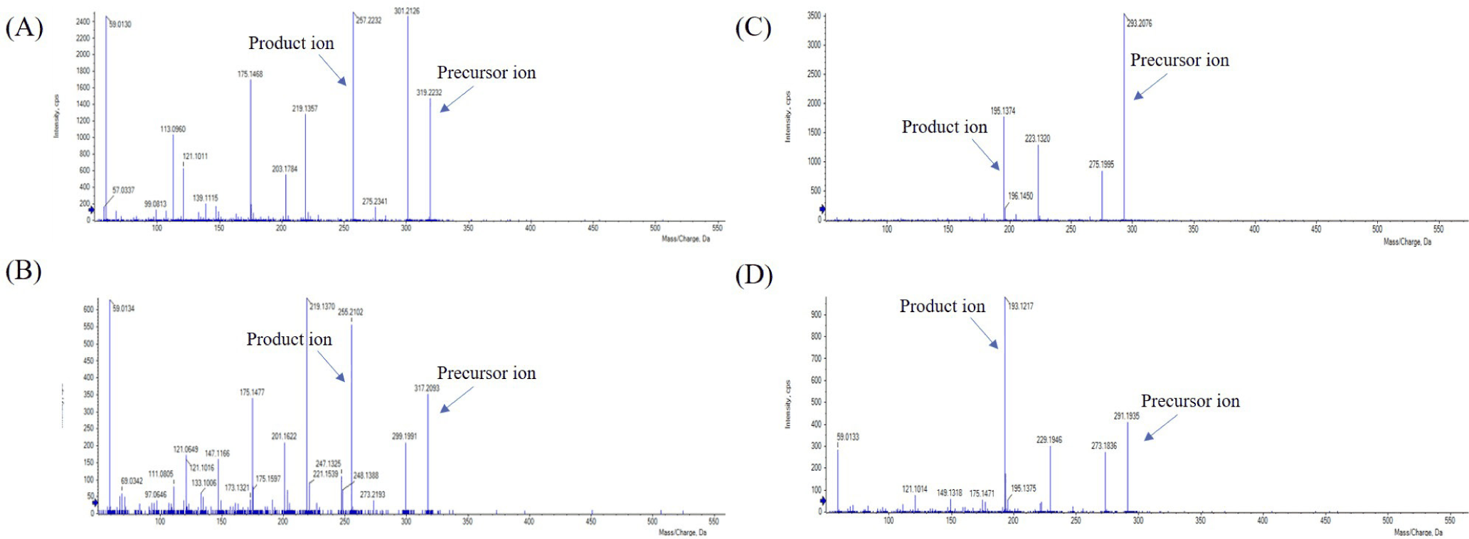
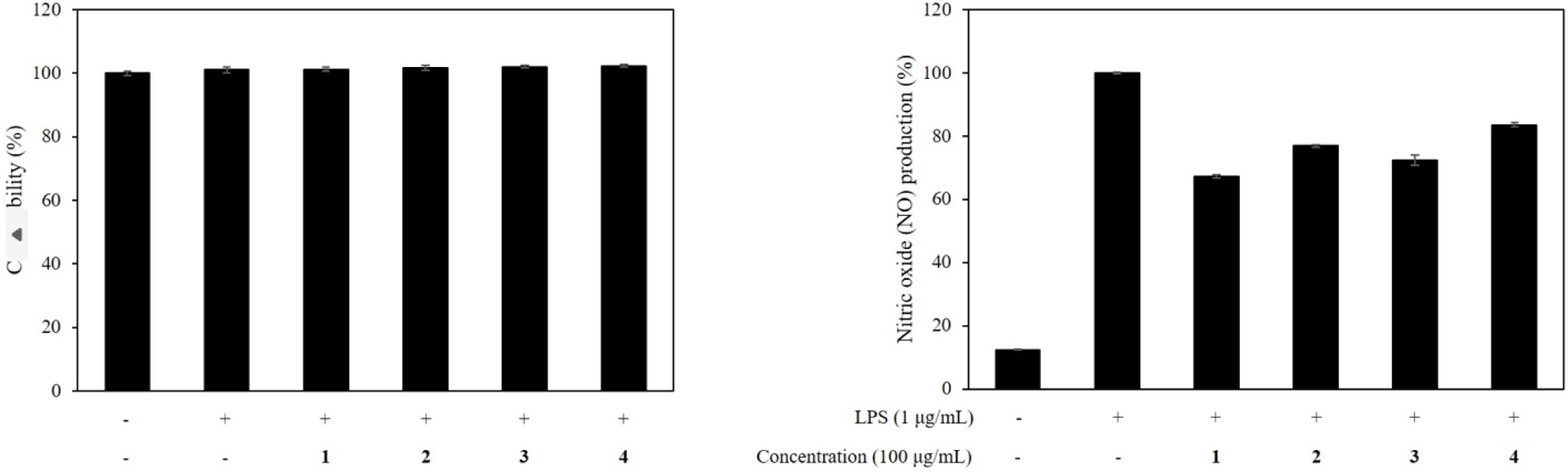
Compound 4 was also obtained as a yellow amorphous gum. Its molecular formula was determined to be C18H28O3 using HR-ESI-MS, based on a negative ion peak at m/z 291.1948 (calcd for C18H27O3, 291.1960). The 1H and 13C NMR spectra (Table 1) of 4 were similar to those of 3 except for the replacement of the double bond at H-6 (δH 5.40)/H-7 (δH 5.37) with single bond at H-6 (δH 1.33)/H-7 (δH 1.40). However, the carbonyl carbon was not detected due to low intensity. The double bond at H-6 (δH 5.40)/H-7 (δH 5.37) was elucidated by HMBC correlations from H-4 (δH 1.42) to C-6 (δC 131.3), from H-5 (δH 2.11) to C-6 (δC 131.3), C-7 (δC 128.8), and from H-8 (δH 2.94) to C-6 (δC 131.3), C-7 (δC 128.8). The 1H and 13C NMR signals at H-2 (δH 2.26)/C-2 (δC 36.3) in 4 suggested the presence of a carbonyl group, in comparison to the proton and carbon NMR signals at δH 2.29/δC 34.6 in 1. The geometries at double bonds were determined to be Z-form (H-6), Z-form (H-9), E-form (H-11), and Z-form (H-15) by comparison of NMR evidence in 1–3. Accordingly, compound 4 was determined to be (6Z,9Z,11E,15Z)-13-hydroxyoctadeca-6,9,11,15-tetraenoic acid. The MS chromatograms were obtained using HR-Q-TOF-MS spectroscopy. Compounds 1–4 were detected at 21.2–22.5 min. The main product ions for compounds 1–4 were delected at m/z 257.2232 [M-H]–, 255.2102 [M-H]–, 195.1374 [M-H]–, and 193.1215 [M-H]– respectively (Fig. 2). The calibration curves and correlation coefficients (R2) are summarized in Table 2. The concentrations of compounds 1–4 in the 70% ethanol extract of E. bicyclis were confirmed to be 0.060 mg/g in 1, 0.155 mg/g in 2, 0.042 mg/g in in 3, and 0.161 mg/g in 4, respectively (Table 2). The cell viability of compounds 1–4 exhibited their non-toxicity at a concentration of 100 mg/mL. We evaluated the inhibitory activity of compounds 1–4 on NO production at the concentrate of 100 mg/mL. The effects of compounds 1–4 on NO production at 100 μg/mL are shown in Fig. 3.
Conclusion
This study describes the isolation, structural identification, and quantification of four known oxylipin derivatives from E. bicyclis. All compounds were determined to be (5Z,8Z,11Z,13E)-15-hydroxyicosa-5,8,11,13-tetraenoic acid (1), (5Z,8Z,11Z,13E,17Z)-15-hydroxyicosa-5,8,11,13,17-pentaenoic acid (2), (9Z,11E,15E)-13-hydroxyoctadeca-9,11,15-trienoic acid (3), and (6Z,9Z,11E,15Z)-13-hydroxyoctadeca-6,9,11,15-tetraenoic acid (4), respectively. Furthermore, four oxylipin derivatives from the 70% EtOH extract of E. bicyclis were evaluated for their anti-inflammatory activities. The results showed the level of NO production were significantly inhibited at the concentration of 100 mg/mL. Quantitative analysis of compounds 1–4 revealed the following contents: 0.060 ± 0.003 mg/g (1), 0.155 ± 0.003 mg/g (2), 0.042 ± 0.003 mg/g (3), and 0.161 ± 0.009 mg/g (4). Phlorotannins have been reported as the major bioactive compounds in the brown alga E. bicyclis. In contrast, the present study demonstrates that oxylipin derivatives also contribute to the biological profile of this species, with the primary focus on the quantitative characterization of four known oxylipins (1–4) in the 70% ethanol extract, accompanied by a preliminary evaluation of their NO inhibitory activity at a concentration of 100 μg/mL. Among the identified oxylipins, compound 4 was found to be the most abundant. Furthermore, oxylipins from E. bicyclis were reported to have anti-microbial activities against two strains. Oxylipins are oxidized polyunsaturated fatty acid derivatives, which has been reported to regulate a wide range of biological functions (Ağagündüz et al., 2024; Barbosa et al., 2016; Misheva et al., 2022). These findings indicate that diverse biological activities of E. bicyclis could be attributed to both phlorotannins and oxylipins. Taken together, these results suggest that the biological activities of E. bicyclis may arise from multiple classes of metabolites, highlighting its potential as a source of anti-inflammatory bioactive compounds.


