

Introduction
From 1961 to 2021, global seafood production experienced an average annual growth rate of 3%. By 2022, total production reached 185 million tonnes, with aquaculture contributing 94 million tonnes (51%), surpassing capture fisheries for the first time (FAO, 2024). This highlights the increasingly vital role of aquaculture in meeting the global demand for seafood. In the Korea, the olive flounder (Paralichthys olivaceus) is a premier aquaculture species. As of 2024, its production reached 40,125 tonnes, accounting for 55% of the total domestic fish aquaculture yield, with an economic value of approximately 684.2 billion KRW (Statistics Korea, 2025).
However, high-density aquaculture environments inevitably elevate the risk of infectious disease outbreaks (Krkošek, 2010), necessitating the continuous use of antibiotics. In 2015, total aquaculture antibiotic usage in Korea was 102,929 kg, of which amoxicillin, a broad-spectrum beta-lactam, was the most frequently utilized (15,594 kg, 15.5%) (Kim et al., 2019). For olive flounder, amoxicillin is typically administered via intramuscular (IM) injection at a dose of 40 mg/kg body weight (BW), which legally requires a 20-day withdrawal period to ensure food safety (NFQS, 2025).
Conventionally, withdrawal periods are established based solely on the depletion of antibiotic residues in edible muscle below acceptable safety thresholds. However, IM injections inherently cause mechanical and chemical tissue damage, leading to local inflammation and necrosis at the injection site (Abbate et al., 2018; Zoltick et al., 2024). If injection-induced lesions persist beyond the withdrawal period, they severely degrade fillet quality. This is particularly problematic in Korea and Japan, where olive flounder is predominantly consumed raw (sashimi). Visible injection marks have previously compromised consumer trust, notably causing a 34% price drop and a sharp decline in exports in Jeju, Korea (Ko, 2023; Park, 2019).
Despite these substantial economic impacts, research evaluating the histopathological recovery at injection sites during the withdrawal period remains scarce. It is currently uncertain whether the statutory 20-day withdrawal period guarantees complete tissue repair, highlighting the urgent need to evaluate alternative, non-edible IM injection sites. A recent pharmacokinetic study by Lee et al. (2023) established that administering amoxicillin via cheek muscle injection yields sufficient systemic serum exposure comparable to the traditional dorsal muscle injection, thereby validating its theoretical therapeutic efficacy against systemic infections. However, critical practical concerns regarding local tissue safety, site-specific absorption kinetics, and the extent of injection-induced histopathological damage have remained entirely unaddressed. Building directly upon the systemic PK framework provided by Lee et al. (2023), this short communication aimed to exclusively evaluate the local histopathological changes and muscle-specific PK characteristics during the withdrawal period following amoxicillin IM injection into both the edible dorsal muscle and the non-edible cheek muscle.
Material and Methods
Olive flounder with an average BW of 408.2 ± 35.7 g were sourced from a private farm in Tongyeong, Gyeongsangnam-do, Korea. Fish weighing over 300 g were specifically selected to ensure sufficient cheek thickness for safe and accurate IM injection into the buccal region. The fish were acclimated for three weeks in 120-L flow-through tanks at 22 ± 0.5°C and fed a commercial pellet diet (CJ Feed, Gunsan, Korea) at 1.5% of BW per day. Prior to drug administration, blood and muscle samples were collected from randomly selected fish to confirm the absence of amoxicillin residues. All experimental procedures were approved by the Institutional Animal Care and Use Committee (IACUC, NFQS-2022-6).
A commercial amoxicillin sodium formulation (AMOX injection, water-soluble powder, 50 g; KBNP Veterinary Medicine, Yesan, Korea) was reconstituted with phosphate-buffered physiological saline to achieve a final dose of 40 mg/kg BW. Intramuscular injections were administered using 1-mL syringes equipped with 3/16" × 23-gauge stainless steel veterinary needles (Socorex Isba SA, Ecublens, Switzerland), reflecting standard aquaculture practices to minimize tissue damage and ensure accurate drug delivery.
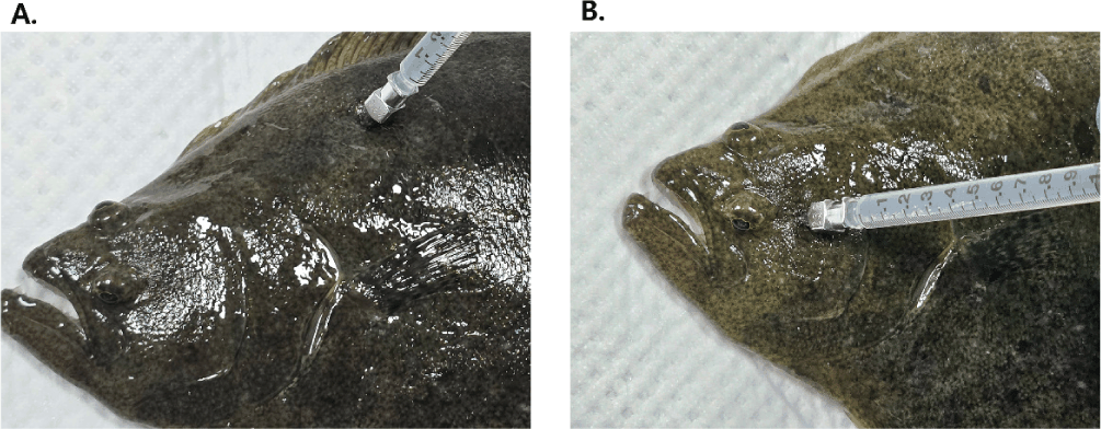
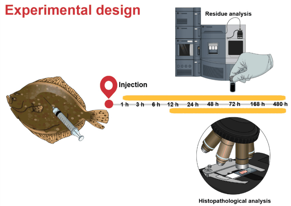
Olive flounder were randomly divided into two groups, each consisting of 70 fish. In a parallel design, one group received a single IM injection of amoxicillin at 40 mg/kg (a dose currently approved in Korea) into the dorsal muscle, while the other group was administered the same dose into the cheek muscle (Fig. 1). Muscle and skin were sampled from seven fish in both groups at each of the 0.5, 1, 3, 6, 12, 24, 48, 72, 168, and 480 hours post-injection (hpi) time points. For these samples, tissue collection was performed after scale removal and included both skin and underlying muscle to reflect edible portions. From the muscle portion of these samples, specific sections measuring 3 × 2 × 1 cm (width × length × depth) were then collected from the injection site for histopathological analysis at 12, 24, 48, 72, 168, and 480 hpi. The overall experimental design is summarized schematically in Fig. 2.
All samples were initially fixed in Bouin’s solution (BBC, Mount Vernon, WA, USA) for 24 hours and then re-fixed in the same solution for 48 hours. The tissues were then immersed in 70% ethanol for 24 hours. Tissue processing—including dehydration, clearing, and paraffin infiltration—was performed using an automatic tissue processor (Leica TP 1020, Wetzlar, Germany). The processed tissues were embedded in paraffin, sectioned into 4–5μm thick slices, and stained using Harris hematoxylin and eosin (H&E) solution (Leica, Teaneck, NJ, USA).
High-performance liquid chromatography (HPLC)-grade water and acetonitrile (J.T. Baker, Phillipsburg, NJ, USA) were used throughout all extraction and chromatographic procedures. Other reagents, including amoxicillin sodium (molecular weight 365.4), piperacillin (molecular weight 517.5), acetic acid, sodium phosphate dibasic (Na2HPO4), potassium dihydrogen phosphate (KH2PO4), and formic acid, were of analytical grade and obtained from Sigma-Aldrich (St. Louis, MO, USA). Solid-phase extraction (SPE) was performed using OASIS PRiME HLB cartridges (6 cc, 200 mg; Waters, Milford, MA, USA). For sample extraction, 2 g of homogenized tissue samples (muscle including skin) were placed into 50-mL conical polypropylene tubes. After adding 1 mL of 0.1 N acetic acid, the samples were subjected to sonication at 60 Hz and 300 W for 5 min at 25°C. Methanol (8 mL) and an internal standard (IS) solution (piperacillin, 500 ppb, 200μL) were subsequently added, followed by centrifugation at 20,000×g for 10 min at 4°C. The supernatant was carefully transferred to 15-mL conical tubes and evaporated under a nitrogen stream at 50°C. The dried residue was reconstituted in 5 mL of 0.1 M Na2HPO4 (pH 7.2) and loaded onto preconditioned SPE cartridges. The cartridges were rinsed with 5 mL of 0.05 M Na2HPO4 and eluted with 5 mL of a 0.05 M KH2PO4 : Acetonitrile mixture (50:50, v/v). After evaporation of the eluate under nitrogen at 50°C, the final extract was dissolved in 1 mL of water and filtered using 0.2-μm polyvinylidene fluoride (PVDF) syringe filters prior to high-performance liquid chromatography–tandem mass spectrometry (HPLC)-MS/MS analysis.
Piperacillin was selected as the internal standard based on the guidelines of the Ministry of Food and Drug Safety (MFDS, 2023). It is aβ-lactam antibiotic with similar extraction and ionization behavior to amoxicillin, and it is entirely absent endogenously in olive flounder tissue. The analytical method was validated with a limit of quantitation (LOQ) of 10 ng/mL for muscle matrices. The matrix-matched calibration curve exhibited excellent linearity across the range of 10–500 ng/mL. Inter-day precision, assessed at 1×, 5×, and 10× LOQ levels, demonstrated a coefficient of variation below 12.12%. Mean recovery rates ranged from 95.44% to 109.92%, ensuring a stable response consistent with the acceptance criteria outlined in the Bioanalytical Method Validation Guidance for Industry. Under the established chromatographic conditions, the retention times were 2.81 min for amoxicillin and 9.88 min for piperacillin (IS), demonstrating successful baseline separation. Detailed HPLC-MS/MS analytical conditions are summarized in Table 1.
Pharmacokinetic parameters of amoxicillin in muscle tissue were determined using non-compartmental analysis (NCA) based on statistical moment theory, conducted with the PKSolver add-in program for Microsoft Excel (Zhang et al., 2010). The maximum concentration (Cmax) and time to reach Cmax (Tmax) were obtained directly from the observed data. The area under the concentration–time curve (AUC) and the area under the first moment curve (AUMC) was calculated using the linear trapezoidal method. The terminal elimination rate constant (λz) was estimated by linear regression of the log-transformed concentration data, and the terminal half-life (t1/2 λz) was calculated as ln 2 divided by λz. The mean residence time (MRT) was determined by dividing AUMC by AUC.
Pharmacodynamic evaluations utilized minimum inhibitory concentration (MIC90) values of 0.0312 µg/mL for Streptococcus iniae and 0.5 µg/mL for Streptococcus parauberis, adopted from published susceptibility datasets for clinical isolates from Korean olive flounder (Lim et al., 2017). Because MIC distributions may vary by region and time, these indices should be interpreted as exposure comparisons under the selected MIC90 scenario. Based on the concentration–time profiles in the muscle, PK/PD exposure indices including Cmax/MIC90, AUC/MIC90, and the duration of time that concentrations exceeded MIC90 (T > MIC90) were calculated. Because the tissue-specific free fraction (fu) of amoxicillin in fish muscle remains empirically undetermined, the total drug concentration was used; therefore, the index is expressed as T > MIC90 (total). Since only the unbound drug is microbiologically active, particularly for time-dependentβ-lactams like amoxicillin, these indices represent the upper bound of potential local antibacterial pressure.
It must be emphasized that no blood samples were collected, and serum PK parameters were not empirically evaluated in the present study. To provide a systemic baseline for therapeutic efficacy, we extracted the established serum PK profiles from a previous foundational study (Lee et al., 2023) conducted under identical dosing and environmental conditions. The PK/PD-related exposure indices in muscle obtained from the present study were calculated strictly to compare local tissue exposure and drug retention between injection sites, providing pharmacological evidence for injection-induced tissue damage, and were not intended to predict the clinical cure of systemic streptococcosis.
Results
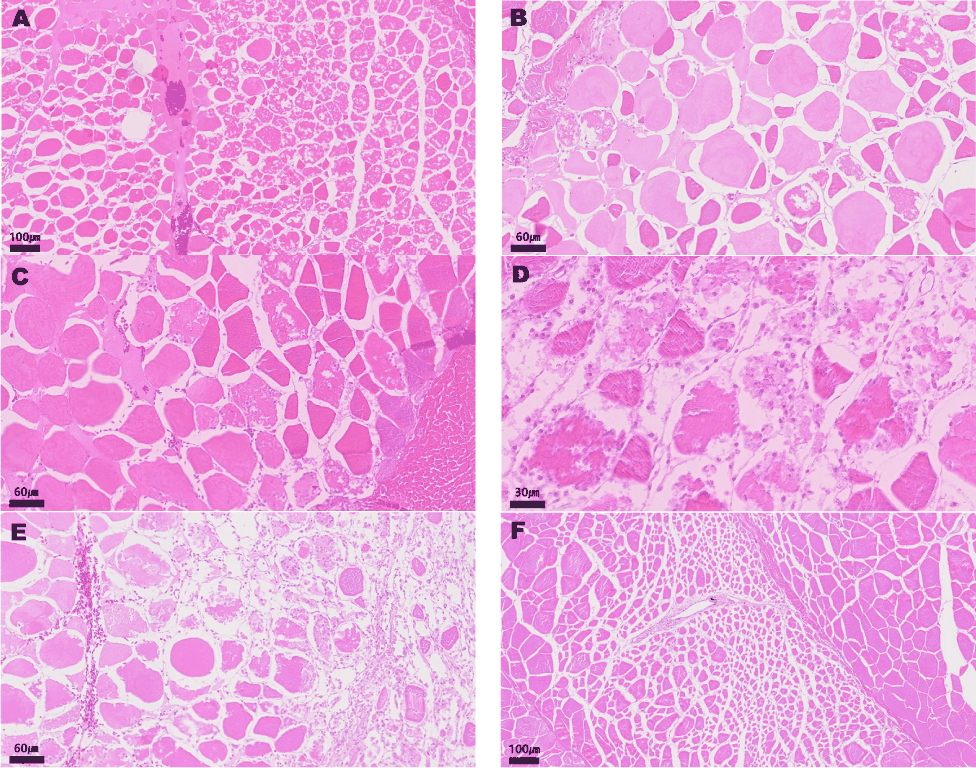
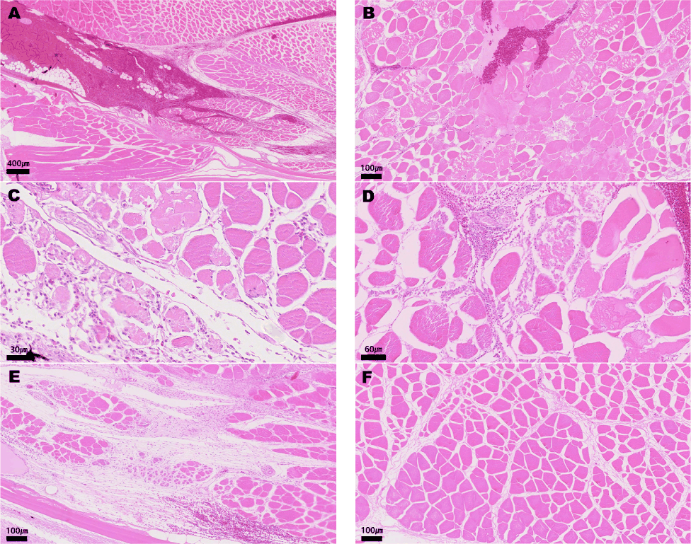
IM injection of amoxicillin induced distinct time-dependent histopathological changes in both the dorsal and cheek muscles at the injection sites. Representative histological lesions were consistently observed in both muscle groups, demonstrating a comparable pattern of tissue damage and recovery over time. At 12 hours post-injection, both dorsal and cheek muscles exhibited extensive hemorrhage, interstitial edema, myofiber atrophy, and necrosis. These findings indicate acute muscle injury occurring shortly after drug administration. By 24 and 48 hours, edema and necrosis remained prominent, with fragmented myofibers and inflammatory cell infiltration becoming evident. At 72 hours, inflammation intensified and was accompanied by partial hyalinization and ongoing fragmentation of muscle fibers, suggesting ongoing tissue degeneration. By 168 hours, widespread and diffuse inflammatory infiltrates persisted in both groups, reflecting a sustained immune response. Even at 480 hours post-injection, histological examination revealed persistent interstitial edema and persistent atrophy of affected myofibers, with only partial resolution of inflammation. Overall, both the dorsal and cheek muscles demonstrated substantial damage following intramuscular administration of amoxicillin. Despite gradual reduction of hemorrhage and inflammatory activity over time, full histological recovery was not achieved within the 20-day observation period (Figs. 3 and 4).
Following intramuscular administration of amoxicillin sodium into the cheek and dorsal muscles of olive flounder, tissue concentrations of the drug were measured at multiple time points using HPLC–MS/MS (Fig. 5). Sampling was performed at 0.5, 1, 3, 6, 12, 24, 48, 72, 168, and 480 hours post-injection, with seven fish analyzed per time point. To visualize inter-individual variation, concentrations from all individuals were plotted as individual data points. Fig. 5A shows the comparative concentration–time profiles between the two injection sites, while Fig. 5B and 5C present individual profiles for the dorsal and cheek muscles, respectively, along with magnified views of the terminal phase, focusing on the decline below the maximum residue limit (MRL; 0.05 mg/kg).
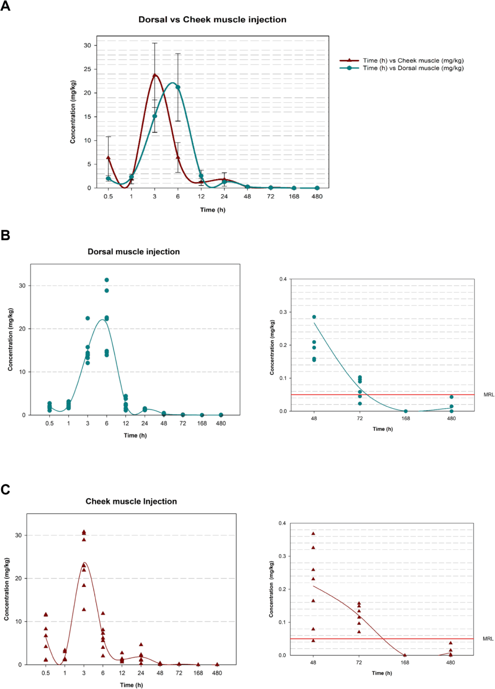
In the cheek muscle group, amoxicillin was rapidly absorbed, reaching the peak concentration (Cmax) at 3 hours, followed by a sharp decline. In some individuals, a secondary rise in concentration was observed. In contrast, the dorsal muscle group exhibited a more gradual increase in drug concentration, with a relatively flattened terminal phase.
The rate at which the drug concentration declined below the MRL was faster in the dorsal muscle group, where most samples fell below the threshold after 168 hours. However, in the cheek muscle group, detectable concentrations near the MRL were still observed in a few individuals at 480 hours. Notably, greater inter-individual variability was observed in the cheek muscle group compared to the dorsal group, indicating higher variability in local absorption and/or distribution dynamics at that site.
Following the IM injection of amoxicillin into the dorsal or cheek muscle of olive flounder, distinct pharmacokinetic profiles were observed in the serum versus muscle compartments (Table 2). It should be noted that direct statistical comparisons between the two compartments were not performed because the serum parameters were derived from the literature (Lee et al., 2023), whereas the muscle parameters were obtained from the present in vivo study. All differences are described in terms of observed trends.
PK/PD indices were calculated using MIC90 values of 0.0312 µg/mL for Streptococcusiniae and 0.5 µg/mL for Streptococcus parauberis.
Cmax, maximum observed concentration; Tmax, time to reach Cmax; AUC₀–t, area under the concentration–time curve from time zero to the last quantifiable concentration; AUMC0–inf, area under the first moment curve extrapolated to infinity; MRT0–inf, mean residence time extrapolated to infinity.
Serum PK parameters were based on data from Lee et al. (2023), while muscle parameters were derived from the present study using non-compartmental analysis.
In the systemic circulation (serum data adapted from Lee et al., 2023), amoxicillin reached a high peak concentration (Cmax) of approximately 203 µg/mL at 3 h post-injection for both the dorsal and cheek injection sites. Systemic exposure, as indicated by AUC0-t, was robust in both groups, confirming that both injection sites provide sufficient systemic bioavailability for treating systemic infections.
In contrast, within the muscle tissue (present study), local drug absorption was generally slower and exhibited different kinetic patterns. The Tmax values were 6 h and 3 h for the dorsal and cheek injections, respectively. The dorsal muscle group showed a higher local Cmax (21.19 µg/mL) and AUC0-t (207.91 µg/mL·h) than the cheek muscle group. Notably, the mean residence time MRT0-inf in the muscle tissue was longer for the cheek injection (31.43 h) compared to the dorsal injection (23.08 h).
To evaluate the extent of local tissue exposure—which correlates with injection-site damage—PK/PD-related indices were calculated based on the total drug concentrations in the muscle. Against S. iniae (MIC90 = 0.0312 µg/mL) and S. parauberis (MIC90 = 0.5 µg/mL), the dorsal injection consistently produced higher local exposure indices (Cmax / MIC90, AUC0-t / MIC90, and T > MIC90 (total)) compared to the cheek injection. These findings indicate that while serum exposure is highly favorable for both sites (Lee et al., 2023), the dorsal muscle experiences a greater magnitude of local drug exposure, providing a pharmacological basis for the observed severe injection-induced tissue damage and the residue depletion patterns during the withdrawal period.
Discussion
IM injection is widely recognized to cause muscle damage, including hemorrhage, inflammation, and tissue necrosis in terrestrial animals such as humans, cattle, and pigs (Abbate et al., 2018; Zoltick et al., 2024). To minimize these adverse effects, international organizations such as the World Health Organization (WHO) and Centers for Disease Control and Prevention (CDC) have issued guidelines on safe injection practices (CDC, 2025; WHO, 2016). Despite these efforts, adverse tissue reactions continue to occur. For instance, in the swine industry, damaged muscle tissue at injection sites is routinely excised during slaughter to ensure food safety. In the Korea, although injection guidelines for farmed fish have been established, the recommended injection site is strictly limited to the anterior dorsal muscle between the dorsal fin and lateral line (Jeong, 2006). Unlike livestock processing, however, the removal of injection-site lesions is not standard practice in the seafood supply chain. Because this traditional dorsal injection site is a primary edible portion, persistent injection marks remain visible to end-consumers. Therefore, this study evaluated the cheek muscle, a non-edible anatomical site, as a practical alternative injection location.
Muscle damage from IM injection typically results from a combination of chemical drug toxicity, physical needle trauma, and the osmotic effects of the injected solution (Svendsen et al., 1998). Tissue recovery generally progresses through four stages: hemostasis, inflammation, proliferation, and remodeling (Velnar et al., 2009), a process that occurs similarly in teleost (Finn & Nielson, 1971). In the present study, all four stages of wound healing were evident at both the dorsal and cheek sites, beginning with initial hemorrhage, followed by extensive inflammation and necrosis, and eventually showing a gradual reduction in the inflammatory response. The recovery trajectories were macroscopically and microscopically similar over time between the two injection sites. However, even after the mandatory 20-day withdrawal period, the tissues remained in the proliferation and remodeling stages, and complete histological recovery was not achieved. This critical finding suggests that current statutory withdrawal periods, which are established solely based on drug elimination kinetics from edible tissues, may be insufficient to ensure complete physiological recovery from injection-induced trauma.
Because the histopathological analysis revealed no significant differences in the severity of tissue damage between the cheek and dorsal muscles, the site-specific muscle residue depletion and pharmacokinetic profiles were comparatively analyzed to understand the local drug dynamics. The analysis revealed distinct kinetic differences: the cheek muscle group reached its maximum local concentration (Cmax) rapidly at 3 hours post-administration, followed by a sharp decline, whereas the dorsal muscle group exhibited a slower rise and a prolonged elimination phase. These differences likely reflect the anatomical and physiological characteristics of the cheek muscle, which possesses a richer vascular supply compared to the dorsal musculature, facilitating more rapid drug absorption and distribution into the systemic circulation. Notably, the cheek muscle group exhibited substantial inter-individual variability, with secondary concentration peaks observed in several individuals. This variability suggests potential drug redistribution or delayed absorption mediated by the specific anatomical confines of the buccal cavity. Furthermore, some individuals in the cheek group maintained detectable concentrations near the MRL up to 480 hours, underscoring the necessity for robust safety assessments that account for high inter-individual variations in alternative injection sites.
As this study was designed as a direct experimental extension of Lee et al. (2023)—utilizing olive flounder from the same farm and batch under identical dosing and environmental conditions—their serum PK/PD data serve as a scientifically robust systemic reference. Regarding the pharmacokinetic/pharmacodynamic (PK/PD) relationships, a clear distinction must be made between systemic therapeutic efficacy and local tissue exposure. As previously established by Lee et al. (2023), cheek muscle injection yields comparable or superior serum PK/PD indices against S. iniae and S. parauberis compared to dorsal injection, ensuring sufficient systemic exposure to eradicate these pathogens. However, our current muscle-specific PK/PD analysis revealed a different dynamic at the local level. The dorsal muscle injection resulted in higher local exposure indices (AUC0-t / MIC90 and T > MIC90) within the muscle tissue itself. This heightened and prolonged local drug retention in the dorsal muscle provides a pharmacological rationale for the sustained tissue damage observed in this study. Thus, while both sites are viable for delivering systemic therapy, transitioning to the non-edible cheek muscle offers the distinct advantage of isolating this inevitable local tissue damage away from the economically valuable fillet.
Several methodological limitations of this study should be acknowledged. First, a sham-injected (PBS/saline) control group was not included. However, a previous study evaluating intramuscular injections in olive flounder demonstrated that the administration of PBS alone does not induce significant or prolonged histopathological alterations at the injection site (Joo et al., 2020). Therefore, while the exact relative contributions of mechanical needle trauma versus amoxicillin-specific cytotoxicity cannot be perfectly isolated, the extensive, severe, and persistent tissue damage observed in our study is highly likely driven primarily by the chemical toxicity of the amoxicillin formulation itself. Second, because the specific free fraction (protein binding rate) of amoxicillin in fish tissue has not been empirically determined, the PK/PD indices (such as T > MIC90) were calculated based on total drug concentrations. As the antibacterial efficacy of beta-lactams is primarily driven by the free drug fraction (fT > MIC) (Cars, 1997), this may overestimate the active drug exposure. If amoxicillin exhibits significant protein or tissue binding in olive flounder muscle, the actual fT > MIC90 would be lower than reported here, potentially overestimating the local antibacterial pressure at the injection site. Furthermore, the degree of overestimation may differ between the dorsal and cheek muscle compartments if tissue-specific binding varies between sites; therefore, the PK/PD indices reported here should be interpreted strictly as relative comparisons of local drug retention rather than absolute predictors of antibacterial efficacy. While the use of a synchronized batch significantly reduces inter-group variability, inherent individual physiological differences in drug absorption and distribution cannot be entirely discounted, which may introduce a degree of imprecision in the direct cross-compartment comparison. Finally, this study was conducted using healthy individuals, which may not fully capture the pharmacokinetic alterations that occur during actual diseased states. Future research should focus on the standardization of alternative injection techniques, evaluation under clinical infection scenarios, and the determination of specific protein binding profiles in teleost.
Conclusion
This study evaluated the clinical applicability of the cheek muscle as an alternative to the conventional dorsal injection site by comparing amoxicillin residue depletion patterns and histopathological changes in olive flounder. As an extension of the systemic pharmacokinetic framework established by Lee et al. (2023), this work elucidates the local tissue dynamics and physical consequences occurring directly at the injection sites. Our findings demonstrate that while the statutory 20-day withdrawal period is sufficient for drug elimination below the MRL, it does not guarantee complete histological recovery from injection-induced trauma. Therefore, transitioning to a non-edible administration site such as the cheek muscle offers a highly practical solution to preserve fillet quality and consumer confidence without compromising systemic therapeutic efficacy. To establish comprehensive, evidence-based recommendations for aquaculture medicine, future investigations must explore standardized injection protocols, multi-species applications, and the pharmacokinetic impacts of diseased states.


