RESEARCH ARTICLE

Hot water extract of Loliolus beka attenuates methylglyoxal-induced advanced glycation end products formation in human umbilical vein endothelial cells

Seon-Heui Cha1,*https://orcid.org/0000-0002-5004-3223, Hee-Sook Jun2,3,4,*https://orcid.org/0000-0002-1166-4932
Author Information & Copyright
1Department of Marine Bio and Medical Sciences, Hanseo University, Seosan 31962, Korea
2Lee Gil Ya Cancer and Diabetes Institute, Gachon University, Incheon 21936, Korea
3College of Pharmacy, Gachon University, Incheon 21936, Korea
4Gachon Medical and Convergence Institute, Gachon Gil Medical Center, Incheon 21565, Korea
*Corresponding author: Seon-Heui Cha, Department of Marine Bio and Medical Sciences, Hanseo University, Seosan 31962, Korea, Tel: +82-41-660-1550, E-mail:sunnycha@hanseo.ac.kr
*Corresponding author: Hee-Sook Jun, College of Pharmacy, Gachon University, Incheon 21936, Korea, Tel: +82-32-899-6056, E-mail:hsjun@gachon.ac.kr

Copyright © 2022 The Korean Society of Fisheries and Aquatic Science. This is an Open Access article distributed under the terms of the Creative Commons Attribution Non-Commercial License (http://creativecommons.org/licenses/by-nc/4.0/) which permits unrestricted non-commercial use, distribution, and reproduction in any medium, provided the original work is properly cited.

Received: Jun 06, 2022; Revised: Aug 18, 2022; Accepted: Sep 14, 2022

Published Online: Oct 31, 2022

Abstract

Over production of methylglyoxal (MGO) a highly reactive dicarbonyl compound, has been associated in progressive diabetes with vascular complication. Therefore, we investigated whether hot water extract of Loliolus beka meat (LBM-HWE) presents a preserve effect against MGO-induced cellular damage in human umbilical vein endothelial cells (HUVECs). The LBM-HWE extract showed to inhibit MGO-induced cytotoxicity. Additionally, the LBM-HWE reduced mRNA expression of pro-inflammatory cytokines, and reduced MGO-induced advanced glycation end product (AGEs) formation. Furthermore, LBM-HWE induced glyoxalase-1 mRNA expression and reduced MGO-induced carbonyl protein formation in HUVECs. The results implicate that LBM-HWE has protective ability against MGO-induced HUVECs toxicity by preventing AGEs formation. In conclusion, LBM-HWE could be used as a potential treatment material for the prevention of vascular complications of diabetes.

Keywords: Diabetes complications; Loliolus beka; Metabolic disease; Methylglyoxal; Glycation

Introduction

Protein glycation, and advanced glycation end products (AGEs) are increased with hyperglycemia and are generated by irreversible reactions between carbohydrates and proteins. AGEs are increased and accumulated in the extracellular matrix of the patient with diabetes (Singh et al., 2014), and induces self-oxidation to produce other reactive intermediates, leading to diabetes development and its complications (Chou et al., 2015; Park & Kim, 2012). An AGEs accumulation in progressive prediabetic stages of disease may contribute to the onset of type 2 diabetes (Stirban et al., 2014). Recently, glycation inhibition has been considered as an effective strategy to slow down human senescence and disease development (Baig et al., 2017). So, there is a growing interest in AGE inhibitors, including a searching bioresources (Nagai et al., 2014).

Indeed, elevation of methylglyoxal (MGO) contributes to the vascular diseases pathogenesis implicated with diabetes complications (Schalkwijk & Stehouwer, 2020). Dysfunction of endothelial cell (EC) implies several vascular diseases, including impairment of ECs barrier functions, vasodilation, disturbances in proliferative activities, tube formation properties, angiogenic properties, synthetic function reduction, and inhibition of white blood cells from adhesion and diapedesis (Cernuda-Morollón & Ridley, 2006; Goligorsky, 2005). ECs control the immune system of the vascular layer and have a critical role in the regulation of the metabolic system (Schalkwijk et al., 2020). Therefore, prevention of diabetes complicated with vascular anormalies is much important for maintaining human health.

Loliolus beka is an edible mollusk that is widely used in several countries particularly in Korea as a food. However, it is not known about the advantageous effects of L. beka on the managing vascular-related diseases (Lee et al., 2019). In current study, therefore, we determined whether extract of L. beka meat protects MGO-induced human umbilical vein endothelial cells (HUVECs) damage.

Materials and Methods

Hot water extract of Loliolus beka meat (LBM-HWE) extract

Loliolus beka were obtained from market and identified by Chonnam National University and washed with tap water to remove salt and other impurities. The water extract prepared according to the previous study (Lee et al., 2019).

Cell maintenance

HUVECs line, was purchased from American Type Culture Collection (ATCC, Manassas, VA, USA). HUVECs were maintained in DMEM (Welgene, Gyeongsan, Korea) containing 10% FBS (Welgene) and maintained in 5% CO2 humidified atmosphere and 95% air at 37°C.

Assessment of cell viability

A cell viability was determined using D-PlusTM CCK (Dongin LS, Seoul, Korea) kit. For the assay, cells were seeded onto 96-well plates with 1 × 10⁴ cells/well. After 16 h hot water extract of Loliolus beka meat (LBM-HWE) and/or MGO were treated to the cells. And, the MGO treated to the cells with the indicated concentrations (0.2, 0.4, 0.8, 1, 2, and 4 mM) for 24 h and then LBM-HWE (125, 250, 500, and 1,000 µg/mL) for 24 h. To determine the protective effects of LBM-HWE, cells were treated with 125, 250 µg/mL LBM-HWE for 1 h, prior to addition of 1 mM MGO for 24 h at 37°C.

Reverse transcription quantitative polymerase chain reaction

To prepare cDNA, we used PrimeScriptTM cDNA synthesis kit (Takara Bio, Shiga, Japan) and total RNA was extracted from the cells using Trizol solution (Takara Bio) prior to cDNA synthesis and all the procedures were performed according to the manufacturer’s instructions and the previous study protocol (Cha et al., 2018). Table 1 listed sequences of primers.

Table 1. Primer sequences
Gene name Sequence 5’-3’
TNF-α Forward GAG ATC AAT CGG CCC GAC TA
Reverse ACA GGG CAA TGA TCC CAA AG
IL1-b Forward CCA CAG ACC TTC CAG GAG AAT G
Reverse GTG CAG TTC AGT GAT CGT ACA GG
IFN-γ Forward GTA GCG GAT AAT GGA ACT CTT TTC TT
Reverse AAT TTG GCT CTG CAT TAT TTT TCT G
Glo-1 Forward ATG CGA CCC AGA GTT ACC AC
Reverse CCA GGC CTT TCA TTT TAC CA
RAGE Forward GTG GGG ACA TGT GTG TCA GAG GGA A
Reverse TGA GGA GAG GGC TGG GCA GGG ACT
Cyclophilin Forward TGC CAT CGC CAA GGA GTA G
Reverse TGC ACA GAC GGT CAC TCA AA

TNF-α, tumor necrosis factor-α; IL1-β, interlukin-1β; IFN-γ, interferon-γ; Glo-1, glyoxalase 1; RAGE, receptor for advanced glycation end product.

Download Excel Table
Determination of advanced glycation end products

To determined AGEs contents the HUVECs were seeded onto 6-well plates as 4 × 10⁶ cells/well, and the cells were incubated with 250 µg/mL of LBM-HWE for 1 h and then further incubated with/without 1 mM MGO (Sigma, St. Luis, MO, USA) for 24 h. The detail procedure followed as described in the previous study (Cha et al., 2018).

Protein carbonyl content

HUVECs (4 × 10⁶) were seeded in each well onto 6-well plates, and the cells were incubated with 250 µg/mL of LBM-HWE for 1 h and then further incubated with/without 1 mM MGO for 24 h. Carbonylation of protein casing oxidative damage was determined using a Assay Kit for Protein Carbonyl Content (Abcam, Cambridge, UK) accoding to the company’s instructions.

Data statistic

All values are presented as means ± SEM. The unpaired t‐test or one‐way analysis of variance (GraphPad Prism software, San Diego, CA, USA) was used for statistical significance calculation.

Results

Hot water extract of Loliolus beka meat attenuates methylglyoxal-induced cytotoxicity

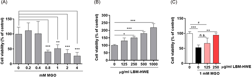
In order to determine whether LBM-HWE has a protective effect on MGO-induced toxicity in human blood vessel cells, HUVECs were treated with either LBM-HWE or MGO alone, or were preincubated with LBM-HWE for 1 h and then further incubated with various doses MGO for various doses. The concentration range tested (125–1,000 µg/mL) of LBM-HWE alone did not show any cytotoxicity in HUVECs, even it induced cell proliferation in dose-dependent manners (Fig. 1B). Cytoviability was significantly lower in MGO-treated HUVECs over 0.8 mM dose (Fig. 1A). Pretreatment with 125, 250 µg/mL LBM-HWE increased the cell viability in dose-dependently, even the viability similar to that of the control at 250 µg/mL LBM-HWE treated cells in the presence of 1 mM MGO for 24 h (Fig. 1C), suggesting that LBM-HWE possesses a protective effect against MGO-induced damage in HUVECs.

fas-25-10-517-g1
Fig. 1. LBM-HWE attenuates MGO-induced cytotoxicity in human umbilical vein endothelial cells. A: The cells were cultured in the presence of 0.2, 0.4, 0.8, 1, 2, and 4 mM MGO for 24 h. B: The cells were cultured with the different concentrations of LBM extract (ext.) for 24 h. C: The cells were incubated with the presence of 0.2, 0.4, 0.8, 1, 2, and 4 mM of MGO for 24 h. A cell viability was subsequently determined. Values were derived from three different experiments. *p < 0.05, **p < 0.01, ***p < 0.001. n.s, no significance; LBM-HWE, Loliolus beka meat hot water extract; MGO, methylglyoxal.
Download Original Figure
Hot water extract of Loliolus beka meat protects against methylglyoxal-induced pro-inflammatory cytokines expression in human umbilical vein endothelial cells

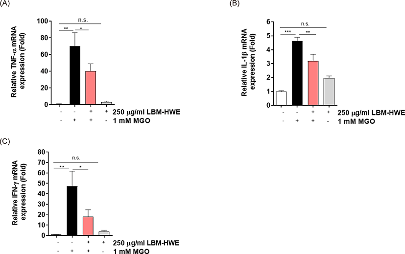
The higher levels (2–4-folds) of MGO is found in diabetic patients plasma (Berlanga et al., 2005; Senda et al., 2015) and exposure of MGO caused inflammation (Berlanga et al., 2005). So, we determined whether LBM-HWE can reduce the production of MGO-induced pro-inflammatory cytokines mRNA expression. As expected, the pro-inflammatory cytokines mRNA levels including IL-1β, TNF-α, and IFN-γ were induced by MGO exposure and its overproduction were reduced as similar to that of the control by LBM-HWE treatment (Fig. 2). These results suggest that LBM-HWE protects MGO-induced pro-inflammatory cytokines activation.

fas-25-10-517-g2
Fig. 2. LBM-HWE attenuates MGO-induced cytotoxicity in human umbilical vein endothelial cells. The cells were cultured in the presence of 250 µg/mL LBM extract (ext.) for 1 h and then further incubated with/without 1 mM MGO for 24 h. Thereafter, the cells were harvested and reverse transcription quantitative polymerase chain reaction was performed for TNF-α (A), IL1-β (B), and IFN-γ (C) mRNA expression. Values were calculated from three independent experiments. *p < 0.05, **p < 0.01, ***p < 0.001. n.s, no significance; TNF-α, tumor necrosis factor-α; IL1-β, interlukin-1β; IFN-γ, interferon-γ; LBM-HWE, Loliolus beka meat hot water extract; MGO, methylglyoxal.
Download Original Figure
Hot water extract of Loliolus beka meat protects against methylglyoxal-induced glycation

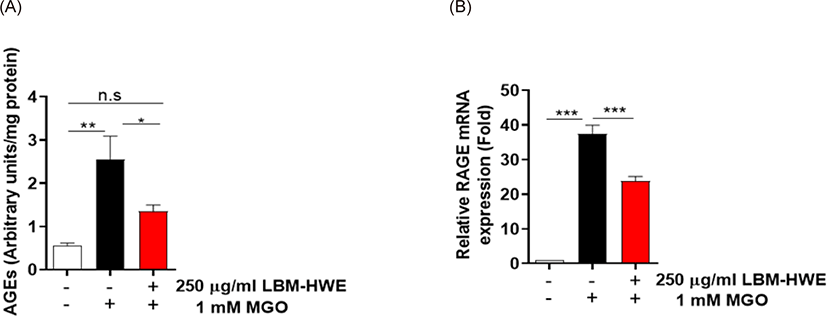
AGE formation is a crucial in complicating with diabetes (Ahmed, 2005). So, we determined whether LBM-HWE reduces AGE formation caused by MGO in HUVECs. A significantly increased (2.5-folds) AGE formation was observed in MGO-treated cells, whereas the induced AGE formation was prohibited by pretreatment with LBM-HWE (Fig. 3A). Due to the harmful effects of AGEs usually interact with its receptor, RAGE (Wang et al., 2019). So, the mRNA expression level of RAGE was also determined. The mRNA expression of RAGE was also significantly induced by MGO exposure, whereas it was reduced by treatment with LBM-HWE prior to MGO (Fig. 3B), suggesting that LBM-HWE induces a protective effect of protein glycation caused by MGO.

fas-25-10-517-g3
Fig. 3. LBM-HWE protects MGO-induced protein glycation in human umbilical vein endothelial cells. The cells were cultured in the presence with 250 µg/mL LBM-HWE extract (ext.) for 1 h and then further incubated with/without 1 mM MGO for 24 h. A: AGE content was measured. B: RAGE mRNA expression level was performed by reverse transcription quantitative polymerase chain reaction. Values were calculated from three independent experiments. *p < 0.05, **p < 0.01, ***p < 0.001. n.s., no significance; LBM-HWE,Loliolus beka meat hot water extract; MGO, methylglyoxal; AGE, advanced glycation end product; RAGE, advanced glycation end product receptor.
Download Original Figure
Hot water extract of Loliolus beka meat protects against methylglyoxal-induced formation of carbonyl proteins

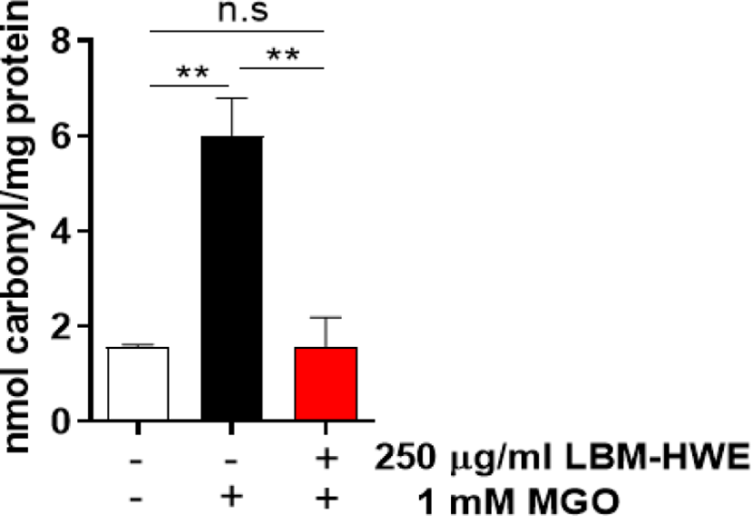
MGO is oxidatively modified to form carbonyl proteins (Allaman et al., 2015; Hanssen et al., 2019). So, we determined whether LBM-HWE protects MGO-induced formation of protein carbonyl, and this content was significantly induced in MGO-treated cells, whereas pretreatment with LBM-HWE prevented MGO-induced protein carbonyl formation (Fig. 4).

fas-25-10-517-g4
Fig. 4. LBM-HWE protects MGO-induced protein glycation in human umbilical vein endothelial cells. The cells were cultured in the presence with 250 µg/mL LBM-HWE for 1 h and then further incubated with/without 1 mM MGO for 24 h. Protein carbonyl content was measured. Values were calculated from three different experiments. **p < 0.01. n.s, no significance; LBM-HWE, Loliolus beka meat hot water extract; MGO, methylglyoxal.
Download Original Figure

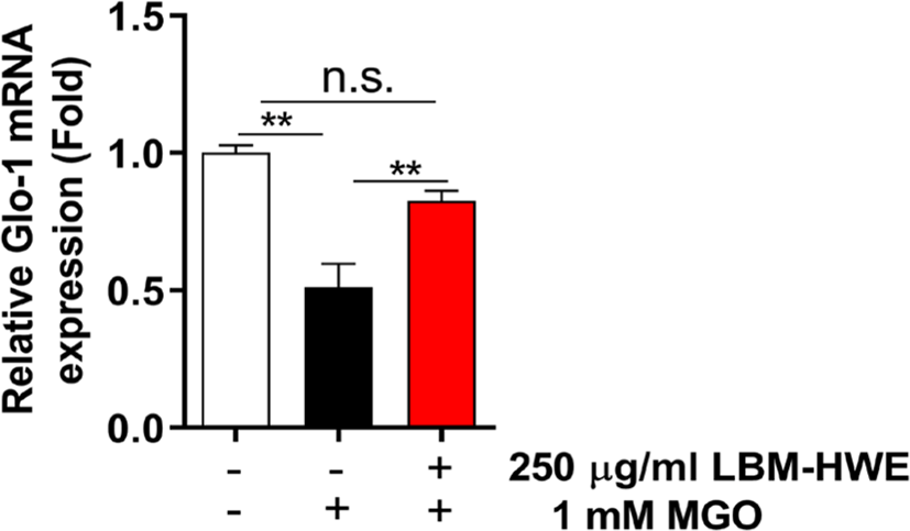
Glycated protein, particularly MGO are detoxified by glyoxalase-1 (Glo-1) (Hanssen et al., 2019). So, we determined glo-1 mRNA expression and found that the expression levels were increased in LBM-HWE-pretreated cells (Fig. 5). These results suggest that LBM-HWE induces a protective effect against MGO-induced protein glycation by increasing the expression of Glo-1 mRNA.

fas-25-10-517-g5
Fig. 5. LBM-HWE extract protects Glo-1 reduction by MGO in human umbilical vein endothelial cells. The cells were incubated with 250 µg/mL LBM-HWE for 1 h and then further incubated with/without 1 mM MGO for 24 h. Glo-1 mRNA expression level was performed by reverse transcription quantitative polymerase chain reaction. Values were derived from three independent experiments. **p < 0.01. n.s., no significance; LBM-HWE, Loliolus beka meat hot water extract; Glo-1, glyoxalase-1; MGO, methylglyoxal.
Download Original Figure

Discussion

A major dicarbonyl compound, MGO, found in higher concentrations of diabetes patients, has been known as a critical player in the development of diabetes complications, including diabetic vascular disease (Cha et al., 2018; Goligorsky, 2005). Recently, in addition, the number of reports is incresing that reactive carbonyls including MGO cause diabetic complications (Pastor-Belda et al., 2017; Senda et al., 2015). Reactive carbonyl responsible for AGE formation is recorded in high concentrations in most patients with diabetes, which is a major factor in the pathogenesis of diabetes mellitus (DM) (Gkogkolou & Böhm, 2012; Kopytek et al., 2020; Pastor-Belda et al., 2017; Senda et al., 2015; Uribarri & He, 2015). Therefore, elimination of accumulated and/or overproduction of AGE or inhibition of its formation is extremely important in treating DM.

Natural products have been emerging as an alternative treatment for diabetes and diabetes complications (Grosso et al., 2017; Heo et al., 2009; Kulkarni, 2014; Xiao & Högger, 2015) in many countries. Loliolus beka is known to rich with taurine and antioxidant effects are found (Han et al., 2017). However, it is not known whether L. beka, might ameliorate diabetic complications. In the present study, we elucidated that the protective effects of hot water extract of L. beka meat (LBM-HWE) on MGO-induced AGE formation in HUVECs.

One of the major causes of the progressive cellular damage in diabetic complications is inflammation (Berlanga et al., 2005). We found that MGO exposure led to toxicity and inflammation and these effects were significantly attenuated by pretreatment with LBM-HWE. A powerful precursor of AGEs, MGO has distinguished as a key signaling molecule mediating pathophysiology of diabetes (Sena et al., 2012). Activation of RAGE induces production of proinflammatory cytokines (Bezold et al., 2019). Administration of MGO to rats causes microvascular changes similar as diabetes symptom (Berlanga et al., 2005), contributing to endothelial (Sena et al., 2012). In our study, MGO exposure induced not only formation of AGEs but RAGE mRNA expression, and this induction reduced by pretreatment with LBM-HWE. Several groups have initiated studies of diabetes etiology and physiology for MGO the management of diabetes complications. Metformin, an antihyperglycemic agent, is popular drug to cure of diabetes patients and known as effective scavenger of MGO by reducing formation of carbonyls (Ruggiero-Lopez et al., 1999).

The glyoxalase (Glo) is a core system for detoxification of MGO and the other reactive aldehydes such as carbonyls (He et al., 2020). Abnormal level of MGO and dysfunction of the Glo system are associated with various diseases, and Glo-1 is an enzyme of Glo system. Our study demonstrated that MGO treatment increased carbonyl protein formation, and this induction was significantly attenuated by pretreatment with LBM-HWE by elevating Glo-1 mRNA expression. Glo-1 prevents hyperglycemia-induced persistent increases DM, which points towards a significant induction of MGO (El-Osta et al., 2008). In addition, it is known that glucose and its metabolites including MGO are accumulated in the cell in diabetic patients (Hanssen et al., 2019). It was reported that when Glo-1 was increased in blood vessel, AGEs production by MGO was inhibited, indicating that MGO acts as a critical mediator for AGE production in vascular cells (Wang et al., 2019), implying that MGO is an AGE precursor that the central key of glycation end products in ECs. In accordance, we demonstrated that induction of Glo-1 by pretreatment of LBM-HWE reduces MGO-induced levels of AGEs in HUVECs. In in vivo data, when Glo-1 decreased, a lower cell viability and higher inflammation-related genes expression were observed in vascular cells (Shinohara et al., 1998). Knockdown of Glo-1 induced MGO concentration and inflammation-related gene expression in human vascular ECs (Stratmann et al., 2016), all these are supporting a role of MGO for endothelial dysfunction. According to Hansen (2001), it has been reported that changes in taurine metabolism led to clinical complications diseases such as retinopathy, neuropathy, nephropathy, cardiomyopathy, platelet aggregation, endothelial dysfunction and atherosclerosis observed in diabetes. The point of view of the treatment of these diabetic complications caused by changes in taurine metabolism, a possible therapeutic view might be to supplement other osmolality and/or small molecule compounds, and taurine in combination therapy with aldose reductase inhibitors. It was confirmed LBM-HWE contained about 40% of taurine (Cha et al., 2019) and had an inhibitory effect on AGE production. There is a previous study that AGE accumulation by MGO directly scavenged overproduced radicals by regulating nrf2 transcription factor (Cha et al., 2019). As well as, LBM-HWE directly scavenged radical in insulin-secreting rat beta cells. There, although, are still only a few studies on the association between AGE accumulation and its inhibition in diabetes, our results proven that the LBM-HWE expected to provide diabetes prevention through the inhibition of AGE accumulation.

Accordingly, LBM-HWE may reduce AGE accumulation caused by MGO, suggesting that LBM-HWE can be used for functional food and pharmaceutical agents related to DM.

Competing interests

No potential conflict of interest relevant to this article was reported.

Funding sources

The Gachon University research fund was supported for this work by of 2019(GCU-2019-0720). We thank to the Basic Science Research Program through the National Research Foundation of Korea (NRF) funded by the Ministry of Science, ICT, & Future Planning (grant number 2020R1C1C1007712).

Acknowledgements

Not applicable.

Availability of data and materials

Not applicable.

Ethics approval and consent to participate

Not applicable.

References

1.

Ahmed N. Advanced glycation endproducts—role in pathology of diabetic complications. Diabetes Res Clin Pract. 2005; 67:3-21

2.

Allaman I, Bélanger M, Magistretti PJ. Methylglyoxal, the dark side of glycolysis. Front Neurosci. 2015; 9-23

3.

Baig MH, Jan AT, Rabbani G, Ahmad K, Ashraf JM, Kim T, et al. Methylglyoxal and advanced glycation end products: insight of the regulatory machinery affecting the myogenic program and of its modulation by natural compounds. Sci Rep. 2017; 7:5916

4.

Berlanga J, Cibrian D, Guillén I, Freyre F, Alba JS, Lopez-Saura P, et al. Methylglyoxal administration induces diabetes-like microvascular changes and perturbs the healing process of cutaneous wounds. Clin Sci. 2005; 109:83-95

5.

Bezold V, Rosenstock P, Scheffler J, Geyer H, Horstkorte R, Bork K. Glycation of macrophages induces expression of pro-inflammatory cytokines and reduces phagocytic efficiency. Aging. 2019; 11:5258-75

6.

Cernuda-Morollón E, Ridley AJ. Rho GTPases and leukocyte adhesion receptor expression and function in endothelial cells. Circ Res. 2006; 98:757-67

7.

Cha SH, Han EJ, Ahn G, Jun HS. Taurine-rich-containing hot water extract of Loliolus beka gray meat scavenges palmitate-induced free radicals and protects against DNA damage in insulin secreting β-cells. Adv Exp Med Biol. 2019; 1155:483-95

8.

Cha SH, Hwang Y, Heo SJ, Jun HS. Diphlorethohydroxycarmalol attenuates methylglyoxal-induced oxidative stress and advanced glycation end product formation in human kidney cells. Oxid Med Cell Longev. 2018; 2018:3654095

9.

Chou CK, Chen SM, Li YC, Huang TC, Lee JA. Low-molecular-weight chitosan scavenges methylglyoxal and Nε-(carboxyethyl)lysine, the major factors contributing to the pathogenesis of nephropathy. SpringerPlus. 2015; 4:312

10.

El-Osta A, Brasacchio D, Yao D, Pocai A, Jones PL, Roeder RG, et al. Transient high glucose causes persistent epigenetic changes and altered gene expression during subsequent normoglycemia. J Exp Med. 2008; 205:2409-17

11.

Gkogkolou P, Böhm M. Advanced glycation end products: key players in skin aging?. Dermato-endocrinol. 2012; 4:259-70

12.

Goligorsky MS. Endothelial cell dysfunction: can’t live with it, how to live without it. Am J Physiol Renal Physiol. 2005; 288:F871-80

13.

Grosso G, Stepaniak U, Micek A, Kozela M, Stefler D, Bobak M, et al. Dietary polyphenol intake and risk of type 2 diabetes in the Polish Arm of the Health, Alcohol and Psychosocial Factors in Eastern Europe (HAPIEE) study. Br J Nutr. 2017; 118:60-8

14.

Han EJ, Um JH, Kim EA, Lee WW, Kang N, Oh JY, et al. Protective effects of an water extracts prepared from Loliolus beka gray meat against H2O2-induced oxidative stress in Chang liver cells and zebrafish embryo model. Adv Exp Med Biol. 2017; 975:585-601

15.

Hansen SH. The role of taurine in diabetes and the development of diabetic complications. Diabetes/Metab Res Rev. 2001; 17:330-46

16.

Hanssen NMJ, Stehouwer CDA, Schalkwijk CG. Methylglyoxal stress, the glyoxalase system, and diabetic chronic kidney disease. Curr Opin Nephrol Hypertens. 2019; 28:26-33

17.

He Y, Zhou C, Huang M, Tang C, Liu X, Yue Y, et al. Glyoxalase system: a systematic review of its biological activity, related-diseases, screening methods and small molecule regulators. Biomed Pharmacother. 2020; 131:110663

18.

Heo SJ, Hwang JY, Choi JI, Han JS, Kim HJ, Jeon YJ. Diphlorethohydroxycarmalol isolated from Ishige okamurae, a brown algae, a potent α-glucosidase and α-amylase inhibitor, alleviates postprandial hyperglycemia in diabetic mice. Eur J Pharmacol. 2009; 615:252-6

19.

Kopytek M, Ząbczyk M, Mazur P, Undas A, Natorska J. Accumulation of advanced glycation end products (AGEs) is associated with the severity of aortic stenosis in patients with concomitant type 2 diabetes. Cardiovasc Diabetol. 2020; 19:92

20.

Kulkarni YA. Diabetes, diabetic complications and natural products. Pharm Crop. 2014; 5:9-10

21.

Lee WW, Han EJ, Shin EJ, Han HJ, Ahn G, Cha SH. Protective effect of hot water extract of Loliolus Beka gray meat against palmitate-induced HUVEC damage. Adv Exp Med Biol. 2019; 1155:717-27

22.

Nagai R, Shirakawa J, Ohno R, Moroishi N, Nagai M. Inhibition of AGEs formation by natural products. Amino Acids. 2014; 46:261-6

23.

Park CH, Kim JW. Effect of advanced glycation end products on oxidative stress and senescence of trabecular meshwork cells. Korean J Ophthalmol. 2012; 26:123-31

24.

Pastor-Belda M, Fernández-García AJ, Campillo N, Pérez-Cárceles MD, Motas M, Hernández-Córdoba M, et al. Glyoxal and methylglyoxal as urinary markers of diabetes. Determination using a dispersive liquid–liquid microextraction procedure combined with gas chromatography–mass spectrometry. J Chromatogr A. 2017; 1509:43-9

25.

Ruggiero-Lopez D, Lecomte M, Moinet G, Patereau G, Lagarde M, Wiernsperger N. Reaction of metformin with dicarbonyl compounds. Possible implication in the inhibition of advanced glycation end product formation. Biochem Pharmacol. 1999; 58:1765-73

26.

Schalkwijk CG, Stehouwer CDA. Methylglyoxal, a highly reactive dicarbonyl compound, in diabetes, its vascular complications, and other age-related diseases. Physiol Rev. 2020; 100:407-61

27.

Sena CM, Matafome P, Crisóstomo J, Rodrigues L, Fernandes R, Pereira P, et al. Methylglyoxal promotes oxidative stress and endothelial dysfunction. Pharmacol Res. 2012; 65:497-506

28.

Senda M, Ogawa S, Nako K, Okamura M, Sakamoto T, Ito S. The strong relation between post-hemodialysis blood methylglyoxal levels and post-hemodialysis blood glucose concentration rise. Clin Exp Nephrol. 2015; 19:527-33

29.

Shinohara M, Thornalley PJ, Giardino I, Beisswenger P, Thorpe SR, Onorato J, et al. Overexpression of glyoxalase-I in bovine endothelial cells inhibits intracellular advanced glycation endproduct formation and prevents hyperglycemia-induced increases in macromolecular endocytosis. J Clin Invest. 1998; 101:1142-7

30.

Singh VP, Bali A, Singh N, Jaggi AS. Advanced glycation end products and diabetic complications. Korean J Physiol Pharmacol. 2014; 18:1-14

31.

Stirban A, Gawlowski T, Roden M. Vascular effects of advanced glycation endproducts: clinical effects and molecular mechanisms. Mol Metab. 2014; 3:94-108

32.

Stratmann B, Engelbrecht B, Espelage BC, Klusmeier N, Tiemann J, Gawlowski T, et al. Glyoxalase 1-knockdown in human aortic endothelial cells – effect on the proteome and endothelial function estimates. Sci Rep. 2016; 6:37737

33.

Uribarri J, He JC. The low AGE diet: a neglected aspect of clinical nephrology practice?. Nephron. 2015; 130:48-53

34.

Wang Z, Zhang J, Chen L, Li J, Zhang H, Guo X. Glycine suppresses AGE/RAGE signaling pathway and subsequent oxidative stress by restoring Glo1 function in the aorta of diabetic rats and in HUVECs. Oxid Med Cell Longev. 2019; 2019:4628962

35.

Xiao JB, Högger P. Dietary polyphenols and type 2 diabetes: current insights and future perspectives. Curr Med Chem. 2015; 22:23-38