RESEARCH ARTICLE

Methanol extract of Myelophycus caespitosus ameliorates oxidative stress-induced cytotoxicity in C2C12 murine myoblasts via activation of heme oxygenase-1

Cheol Park1https://orcid.org/0000-0003-4906-0410, Hyun Hwangbo2,3https://orcid.org/0000-0003-2180-1205, Min Ho Han4https://orcid.org/0000-0002-4293-7630, Jin-Woo Jeong5https://orcid.org/0000-0002-7549-4514, Suengmok Cho6https://orcid.org/0000-0001-5951-5832, Gi-Young Kim7https://orcid.org/0000-0002-6878-0790, Hye-Jin Hwang8https://orcid.org/0000-0002-5448-6443, Yung Hyun Choi2,3,9,*https://orcid.org/0000-0002-1454-3124
Author Information & Copyright
1Division of Basic Sciences, College of Liberal Studies, Dong-Eui University, Busan 47340, Korea
2Department of Biochemistry, Dong-Eui University College of Korean Medicine, Busan 47227, Korea
3Anti-Aging Research Center, Dong-Eui University, Busan 47340, Korea
4National Marine Biodiversity Institute of Korea, Seocheon 33662, Korea
5Honam National Institute of Biological Resources, Mokpo 58762, Korea
6Department of Food Science and Technology, Institute of Food Science, Pukyong National University, Busan 48513, Korea
7Department of Marine Life Science, Jeju National University, Jeju 63243, Korea
8Department of Food and Nutrition, College of Nursing, Healthcare Sciences & Human Ecology, Dong-Eui University, Busan 47340, Korea
9Core-Facility Center for Tissue Regeneration, Dong-Eui University, Busan 47340, Korea
*Corresponding author: Yung Hyun Choi, Department of Biochemistry, Dong-Eui University College of Korean Medicine, Busan 47227, Korea, Tel: +82-51-890-3319, Fax: +82-51-890-3333, E-mail:choiyh@deu.ac.kr

Copyright © 2022 The Korean Society of Fisheries and Aquatic Science. This is an Open Access article distributed under the terms of the Creative Commons Attribution Non-Commercial License (http://creativecommons.org/licenses/by-nc/4.0/) which permits unrestricted non-commercial use, distribution, and reproduction in any medium, provided the original work is properly cited.

Received: Nov 02, 2022; Revised: Nov 15, 2022; Accepted: Nov 15, 2022

Published Online: Jan 31, 2023

Abstract

Myelophycus caespitosus, a brown alga belonging to genus Myelophycus, has been traditionally used as a food and medicinal resource in Northeastern Asia. However, few studies have been conducted on its pharmacological activity. In this study, we evaluated whether methanol extract of M. caespitosus (MEMC) could protect against oxidative damage caused by hydrogen peroxide (H2O2) in C2C12 murine myoblasts. Our results revealed that MEMC could suppress H2O2-induced growth inhibition and DNA damage while blocking the production of reactive oxygen species. In H2O2-treated cells, cell cycle progression was halted at the G2/M phase, accompanied by changes in expression of key cell cycle regulators. However, these effects were attenuated by MEMC. In addition, we found that MEMC protected cells from induction of apoptosis associated with mitochondrial impairment caused by H2O2 treatment. Furthermore, MEMC enhanced the phosphorylation of nuclear factor-erythroid-2 related factor 2 (Nrf2) and expression and activity of heme oxygenase-1 (HO-1) in H2O2-treaetd C2C12 myoblasts. However, such anti-apoptotic and cytoprotective effects of MEMC were greatly abolished by HO-1 inhibitor, suggesting that MEMC could increase Nrf2-mediated activity of HO-1 to protect C2C12 myoblasts from oxidative stress.

Keywords: Myelophycus caespitosus; Reactive oxygen species; DNA damage; Apoptosis; Nuclear factor-erythroid-2 related factor 2 (Nrf2)/heme oxygenase-1 (HO-1)

Introduction

Muscles consume higher amounts of oxygen than other organs to generate energy for muscle contraction. Therefore, they are vulnerable to oxidative stress, in which free radicals accumulate abnormally and excessively (Di Meo et al., 2017; Steinbacher & Eckl, 2015). Muscle cells are differentiated from myoblasts, which are embryonic precursors, through myogenesis process that fuses into multinucleated myotubes (Abrigo et al., 2019; Sambasivan & Tajbakhsh, 2015). Adequate levels of reactive oxygen species (ROS) can regulate cellular signaling pathways required for muscle differentiation, but excessive ROS accumulation is closely associated with impaired muscle formation. In addition, myoblast apoptosis caused by excessive ROS production is accompanied by cell cycle arrest and DNA damage, contributing to blockage of muscle differentiation and induction of muscle atrophy (Santa-Gonzalez et al., 2016; Yu et al., 2016). Therefore, the level of ROS must be tightly controlled to protect muscle function.

Recently, marine resources are receiving great attention as sources of functional foods and nutraceuticals. Among them, it has been reported that extracts or components of edible seaweed can inhibit oxidative stress-dependent pathological conditions such as inflammation, liver damage, dyslipidemia, endothelial dysfunction, and atherosclerosis (Carroll et al., 2021; Pradhan et al., 2020). Their antioxidant activities mainly involve ROS scavenging and activation of intracellular antioxidant signaling pathways. Several previous results have suggested that nuclear factor-erythroid-2 related factor 2 (Nrf2) is critically involved in the antioxidant efficacy of marine algae extracts or ingredients (Besednova et al., 2022; Jayawardena et al., 2018; Lekshmi et al., 2019; Liu et al., 2021). According to our previous studies, ethanol extracts of marine brown algae Sargassum serratifolium and Petalonia binghamiae can suppress DNA damage and apoptotic cell death in oxidative stress-exposed SW1353 human chondrocytes and mouse-derived C2C12 murine myoblasts by Nrf2-dependent activation of heme oxygenase-1 (HO-1) (Kang et al., 2015; Park et al., 2018). Jayawardena et al. (2018) also demonstrated that activation of Nrf2/HO-1 signaling pathway is mediated by the blocking of fine dust-induced oxidative stress by ethanol extract of S. serratifolium in murine macrophages. In addition, it is reported that various components including seaweed-derived polysaccharides and glycoproteins also exhibit antioxidant activity through a similar mechanism (Lekshmi et al., 2019; Niu et al., 2020).

Myelophycus caespitosus Kjellman 1893, a brown alga belonging to genus Myelophycus, is widely distributed along the coast of Northeastern Asia. It has traditionally been used as a food and medicinal resource (Hanyuda et al., 2020). Although we have reported that a methanol extract of M. caespitosus (MEMC) has potent anti-inflammatory effects in BV2 microglial cells (Jayasooriya et al., 2012), scientific evidence for the pharmacological efficacy of M. caespitosus, including its antioxidant properties, is very limited. Therefore, the objective of the present study was to investigate the antioxidant capacity of MEMC against oxidative stress-mediated cytotoxicity in muscle cells. For this purpose, C2C12 mouse myoblasts were used and treated with hydrogen peroxide (H2O2) to mimic oxidative stress.

Materials and Methods

Cell culture

C2C12 cells (CRL-1772TM, ATCC, Manassas, VA, USA) were cultured using the same method as described previously (Park et al., 2021) and used up until passage number 8. All materials necessary for cell culture were purchased from WELGENE (Gyeongsan, Korea). MEMC provided by Jeju Technopark (Jeju, Korea) and H2O2 (Sigma-Aldrich, St. Louis, MO, USA) were dissolved in dimethyl sulfoxide (Thermo Fisher Scientific, Waltham, MA, USA) to make stock solutions. They were used to treat cells after diluting them to appropriate concentrations with culture medium. C2C12 cells were pretreated with or without MEMC, N-acetyl-L-cysteine (NAC, Thermo Fisher Scientific) or zinc protoporphyrin IX (ZnPP, Sigma-Aldrich) for 1 h before they were treated with H2O2 for 24 h or 1 h.

Cell viability assay

To investigate cell viability, 3-(4,5-dimethylthiazol-2-yl)-2,5-diphenyltetra-zolium bromide (MTT, Sigma-Aldrich) assay was applied according to a previously described method (Jeong et al., 2022). Cell morphological changes were observed with an optical microscope (Olympus, Tokyo, Japan).

Flow cytometry analysis

To investigate cell cycle distribution of cells cultured under different conditions, collected cells were fixed with 70% ethanol. As previously described (Choi, 2022), after staining the cells with propidium iodide (PI, Becton Dickinson, San Jose, CA, USA), the frequency of cells corresponding to each cell cycle was calculated using a flow cytometer (Becton Dickinson). For quantitative evaluation of ROS generation, cells were stained with 10 μM 2’,7’-dichlorofluorescein diacetate (DCF-DA, Becton Dickinson) and then DCF fluorescence intensity reflecting ROS generation was analyzed by flow cytometry. To investigate the mitochondrial membrane potential (MMP) using 5,5’,6,6’-tetrachloro-1,1’3,3’-tetraethyl-imidacarbocyanune iodide (JC-1), cells were stained with 2 μM JC-1 (Thermo Fisher Scientific) and then the percentage of JC-1 monomers using a flow cytometer was expressed to indicate cells that lost MMP. For quantitative evaluation of apoptosis-induced cells, cells were stained with annexin V-fluorescein isothiocyanate and PI (Abcam, Cambridge, UK), and then annexin V-positive cells were considered apoptosis-induced cells as described previously (Choi, 2022).

Protein isolation and immunoblotting

Immunoblotting was performed using whole, cytoplasmic and mitochondrial proteins extracted as previously described (Mukherjee et al., 2022). Primary and horseradish peroxidase-conjugated secondary antibodies were obtained from Santa Cruz Biotechnology (Santa Cruz, CA, USA), Cell Signaling Technology (Beverly, MA, USA), or Thermo Fisher Scientific. Information on the primary antibodies used in this study is presented in the Table 1. Cytochrome oxidase subunit 4 and actin were used as loading controls for mitochondrial and cytoplasmic proteins.

Table 1. List of antibodies used in this study
Antibody Species raised Dilution Product code Source
p21 Mouse monoclonal 1:1,000 sc-6246 Santa Cruz Biotechnology
Cyclin A Mouse monoclonal 1:1,000 sc-271682 Santa Cruz Biotechnology
Cyclin B1 Mouse monoclonal 1:1,000 MA5-14319 Thermo Fisher Scientific
Cdk2 Mouse monoclonal 1:1,000 #2546 Cell Signaling Technology
Cdc2 Mouse monoclonal 1:1,000 sc-6248 Santa Cruz Biotechnology
γH2AX Rabbit polyclonal 1:500 PA1-41004 Thermo Fisher Scientific
p-γH2AX Mouse monoclonal 1:500 MA1-2022 Thermo Fisher Scientific
Cytochrome c Mouse monoclonal 1:1,000 sc-13560 Santa Cruz Biotechnology
Bcl-2 Mouse monoclonal 1:1,000 sc-509 Santa Cruz Biotechnology
Bax Mouse monoclonal 1:1,000 sc-7480 Santa Cruz Biotechnology
PARP Mouse monoclonal 1:1,000 sc-8007 Santa Cruz Biotechnology
Nrf2 Mouse monoclonal 1:1,000 sc-518036 Santa Cruz Biotechnology
p-Nrf2 Rabbit polyclonal 1:500 PA5-67520 Thermo Fisher Scientific
HO-1 Mouse monoclonal 1:1,000 sc-136960 Santa Cruz Biotechnology
Keap1 Rabbit polyclonal 1:1,000 #8047 Cell Signaling Technology
COX IV Rabbit polyclonal 1:1,000 #4844 Cell Signaling Technology
Actin Mouse monoclonal 1:1,000 sc-47778 Santa Cruz Biotechnology

Cdk, cyclin-dependent kinase; p-γH2AX, phosphorylation of γH2AX; PARP, poly(adenosine diphosphate-ribose) polymerase; Nrf2, nuclear factor-erythroid-2 related factor 2; p-Nrf2, phosphorylation of Nrf2; HO-1, heme oxygenase-1; Keap1, Kelch-like ECH-associated protein-1; COX IV, cytochrome oxidase subunit 4.

Download Excel Table
Comet assay and nuclear staining

Analysis of DNA damage and morphological identification of apoptotic nuclei were performed through Comet assay kit (Trevigen, Gaithersburg, MD, USA) and 4,6-diamidino-2-phenolindole (DAPI, Sigma-Aldrich) staining (Choi, 2022). After comet assay and DAPI staining, fluorescence images were examined and captured by fluorescence microscopy (Carl Zeiss, Oberkochen, Germany) at Core-Facility Center for Tissue Regeneration, Dong-Eui University.

Heme oxygenase-1 (HO-1) activity assay

To measure HO-1 activity, the amount of bilirubin formed from heme was evaluated using the HO-1 ELISA kit (Abcam). The activity of HO-1 was expressed as fold change relative to the control according to the manufacturer’s instructions.

Statistical analysis

All statistical analyses were performed using GraphPad Prism (Ver. 5.0) (Graphpad, San Diego, CA, USA). Data are expressed as mean ± SD. Statistical differences were determined by one-way analysis of variance with Tukey’s test. Statistical significance was set at p < 0.05.

Results

Methanol extract of Myelophycus caespitosus (MEMC) restores the decrease of cell viability caused by hydrogen peroxide (H2O2)

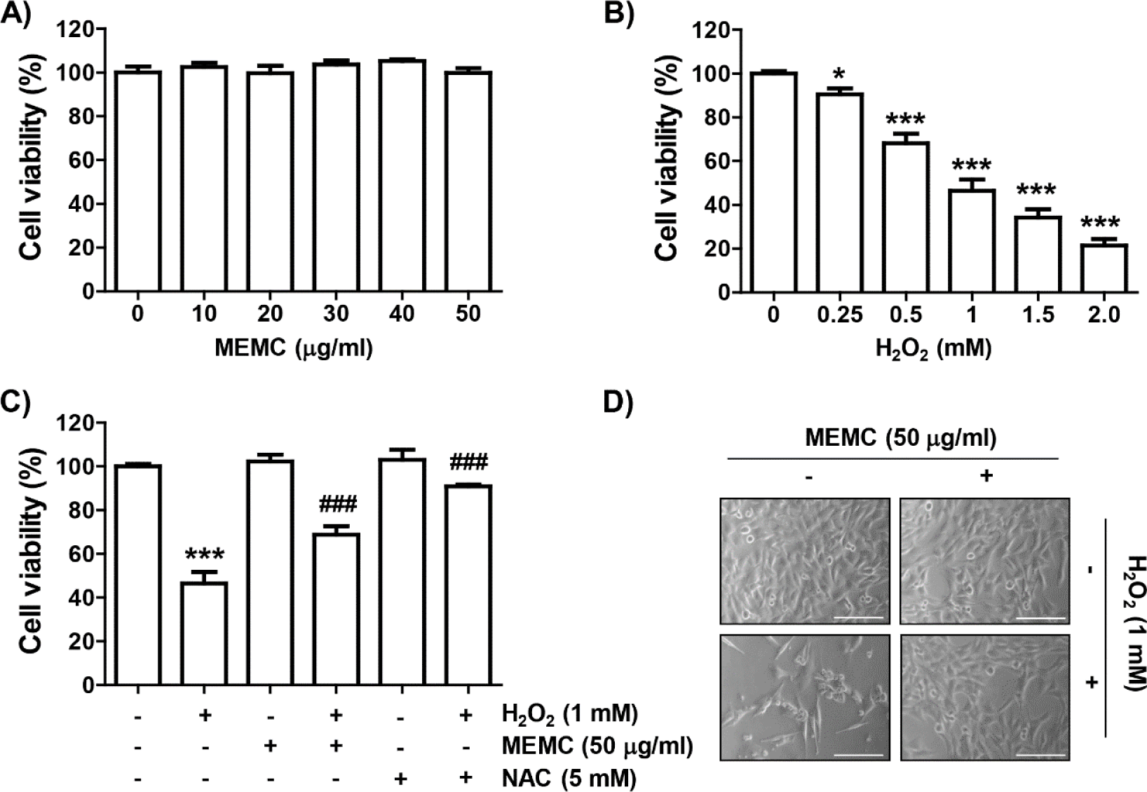
To evaluate the inhibitory activity of MEMC on H2O2-mediated oxidative stress, we assessed investigated the effect of MEMC and H2O2 alone treatment on cell viability of C2C12 cells using the MTT assay. As shown in Fig. 1A, MEMC showed no significant difference in cell viability at concentrations up to 50 mg/mL. Therefore, we used 50 μg/mL of MEMC as the optimal concentration. In H2O2-treated cells, the cell viability was decreased in a dose-dependent manner, and 1 mM H2O2, which reduced cell viability by about half, was set as the cytotoxicity-inducing concentration (Fig. 1B). We next investigated the inhibitory effect of MEMC on H2O2-mediated cytotoxicity and found that MEMC significantly restored H2O2-induced reduction of cell viability and morphological changes (Fig. 1C and 1D), indicating that MEMC pretreatment could improve H2O2-induced cytotoxicity. Furthermore, in the presence of the free radical scavenger NAC, cytotoxicity by H2O2 was completely inhibited (Fig. 1C).
fas-26-1-35-g1
Fig. 1. MEMC ameliorates the reduction of cell viability by H2O2 treatment in C2C12 cells. (A–C) MTT assay was performed on cells that had been cultured under various conditions (*p < 0.05 and ***p < 0.001 vs. control group; ###p < 0.001 vs. H2O2-treated cells). (D) Representative morphological images of cells exposed to H2O2 in the presence or absence of MEMC were presented (× 200). MEMC, methanol extract of Myelophycus caespitosus; H2O2, hydrogen peroxide; NAC, N-acetyl-L-cysteine; MTT, 3-(4,5-dimethylthiazol-2-yl)-2,5-diphenyltetra-zolium bromide.
Download Original Figure
Methanol extract of Myelophycus caespitosus (MEMC) reveres hydrogen peroxide (H2O2)-induced cell cycle arrest

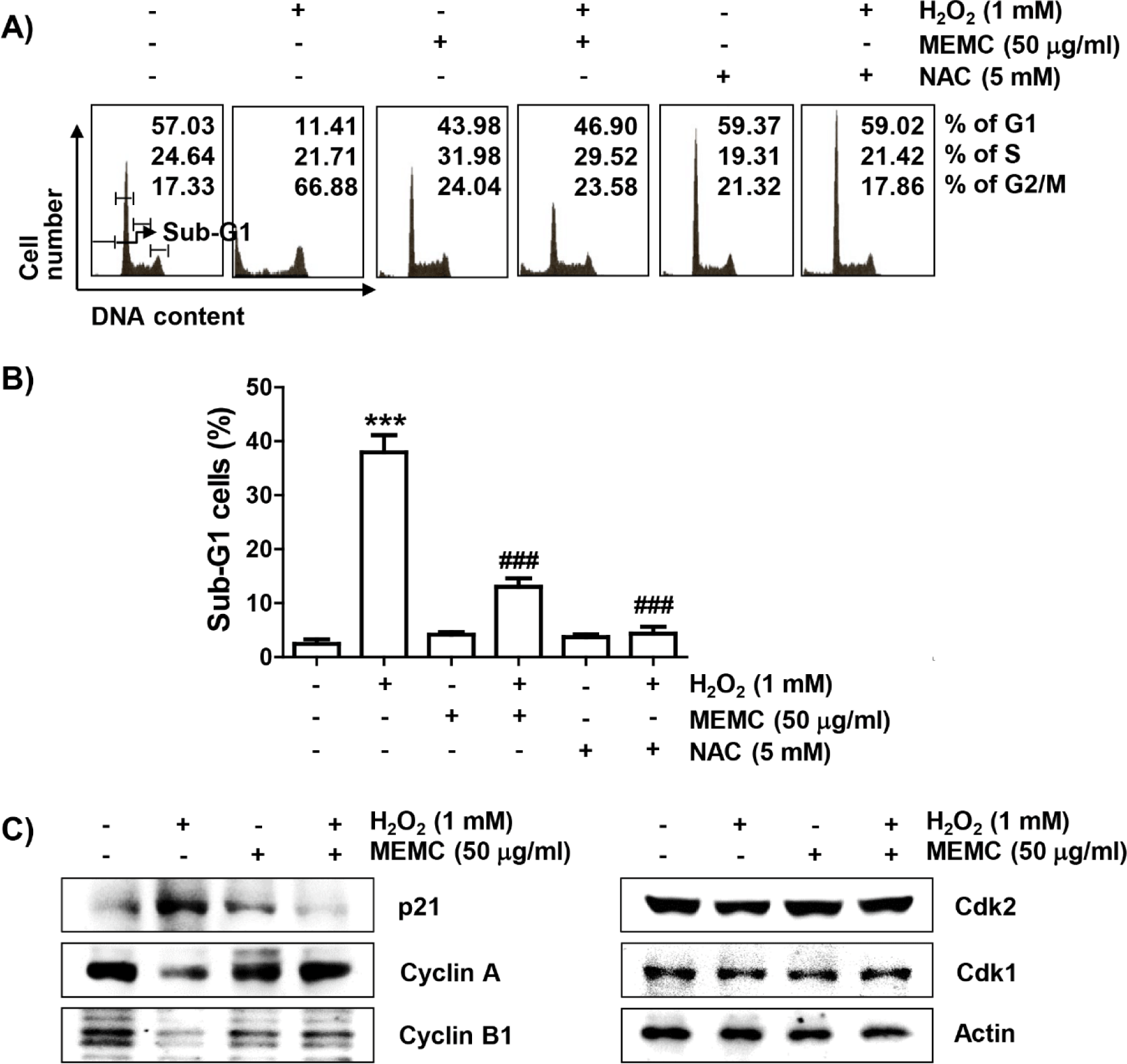
Subsequently, we investigated whether the loss of cell viability in C2C12 cells by H2O2 treatment was due to disturbance of cell cycle progression and induction of apoptosis. As demonstrated in Fig. 2A and 2B, frequencies of cells belonging to G2/M phase and apoptotic sub-G1 phase were increased by H2O2 treatment, but suppressed in the presence of MEMC or NAC. In addition, the expression of p21, protein, one of the representative cyclin-dependent kinase (Cdk) inhibitors, was upregulated in H2O2-treated cells. However, the expression level of cyclin A and cyclin B1 wase suppressed whereas Cdk2 and Cdk1 showed no changes in their expression levels in H2O2-treated cells (Fig. 2C). However, H2O2-induced upregulation of p21 expression was markedly reduced by MEMC treatment, whereas H2O2-induced downregulation of cyclin A and cyclin B1 was inversely increased. These results indicate that the decrease in C2C12 cell survival exposed to H2O2 was due to apoptosis accompanied by cell cycle arrest in the G2/M phase, which was effectively inhibited by MEMC.
fas-26-1-35-g2
Fig. 2. MEMC inhibits H2O2-induced cell cycle arrest in C2C12 cells. Frequencies of cells belonging to each phase (A) and sub-G1 phase (B) of the cell cycle were shown (***p < 0.001 vs. control group; ###p < 0.001 vs. H2O2-treated cells). (C) After extracting cell lysate of each treatment group, the levels of the presented proteins were investigated through immunoblotting. MEMC, methanol extract of Myelophycus caespitosus; H2O2, hydrogen peroxide; NAC, N-acetyl-L-cysteine; Cdk, cyclin-dependent kinase.
Download Original Figure
Methanol extract of Myelophycus caespitosus (MEMC) abolishes reactive oxygen species (ROS) accumulation and DNA damage induced by hydrogen peroxide (H2O2) treatment

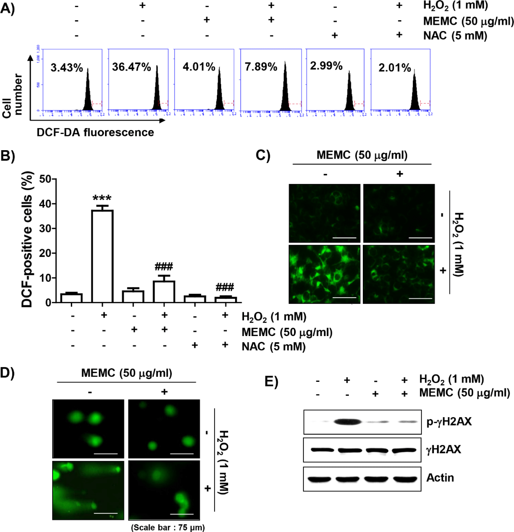
To determine whether the blocking ability of MEMC against H2O2-mediated cytotoxicity was directly related to antioxidant activity, we examined the effect of MEMC on H2O2-induced ROS production using DCF-DA staining. Flow cytometry analysis indicated that ROS production was increased by about 10-fold in C2C12 cells treated with H2O2 for 1 h compared to that in untreated control cells, whereas it was significantly decreased by pretreatment of NAC or MEMC (Fig. 3A, 3B and 3C). Furthermore, in cells treated with H2O2, increases of comet tail moment (DNA migration) and phosphorylation of γH2AX (p-γH2AX) as DNA damage indicators were observed (Fig. 3D and 3E). However, increased DNA migration and phosphorylation of γH2AX by H2O2 treatment were weakened in the presence of MEMC, suggesting that oxidative damage to DNA could be protected by MEMC.
fas-26-1-35-g3
Fig. 3. MEMC suppresses ROS production and DNA damage in H2O2-treated C2C12 cells. Representative flow cytometry histograms examining the extent of ROS generation (A) and analysis results (B) were shown (***p < 0.001 vs. control group; ###p < 0.001 vs. H2O2-treated cells). (C) Representative DCF-DA immunofluorescence images were indicated. (D) Representative immunofluorescence images following comet assay are shown. (E) Changes in expression of γH2AX and its p-γH2AX were determined by immunoblotting. MEMC, methanol extract of Myelophycus caespitosus; H2O2, hydrogen peroxide; NAC, N-acetyl-L-cysteine; p-γH2AX, phosphorylation of γH2AX; ROS, reactive oxygen species; DCF-DA, 2’,7’-dichlorofluorescein diacetate.
Download Original Figure
Methanol extract of Myelophycus caespitosus (MEMC) protects hydrogen peroxide (H2O2)-induced mitochondrial impairment

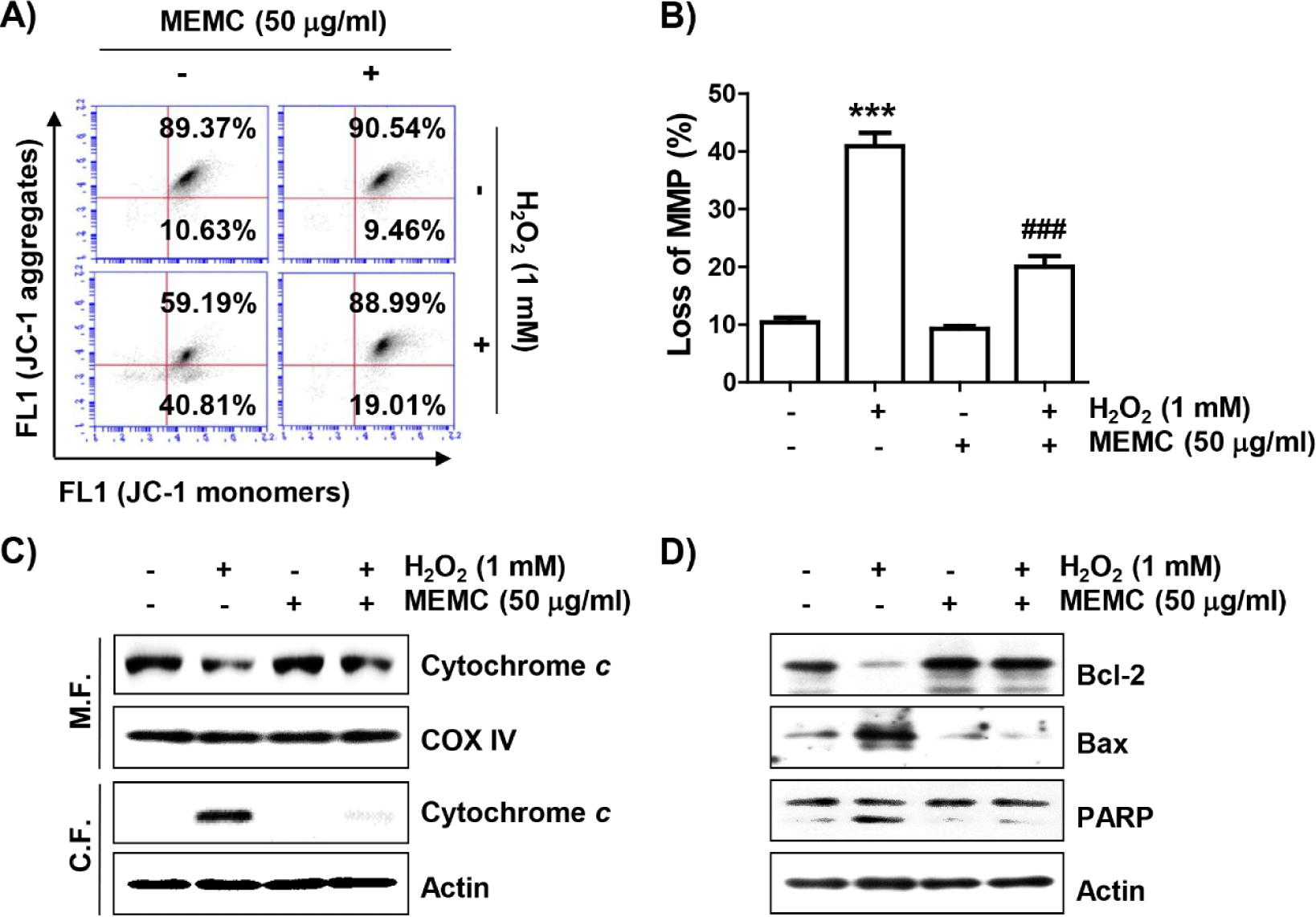
To evaluate whether MEMC could protect against H2O2-induced mitochondrial damage, we estimated MMP following JC-1 staining. JC-1 dye is widely used in apoptosis studies to monitor mitochondrial health, where mitochondria form a monomeric state upon depolarization (Sivandzade et al., 2019). Flow cytometry analysis results (Fig. 4A and 4B) revealed that the frequency of JC-1 monomers was significantly increased whereas that of JC-1 aggregates was decreased in H2O2-treated cells, demonstrating loss of MMP and resultant mitochondrial dysfunction. In addition, after treatment with H2O2, the expression of cytochrome c was increased in the cytoplasmic fraction but decreased in the mitochondrial fraction (Fig. 4C). As shown in Fig. 4D, H2O2 treatment also decreased the expression of Bcl-2 and increased the expression of Bax along with cleavage of poly(adenosine diphosphate-ribose) polymerase (PARP). However, these changes did not occur in cells pretreated with MEMC, suggesting that MEMC was able to inhibit mitochondrial damage caused by H2O2.
fas-26-1-35-g4
Fig. 4. MEMC attenuates H2O2-induced mitochondrial impairment in C2C12 cells. (A) Representative flow cytometry results according to JC-1 staining are presented. (B) The JC-1 monomer ratio of cells in each treatment group was presented (***p < 0.001 vs. control group; ###p < 0.001 vs. H2O2-treated cells). (C and D) The levels of the presented proteins were investigated through immunoblotting. MEMC, methanol extract of Myelophycus caespitosus; H2O2, hydrogen peroxide; JC-1, 5,5’,6,6’-tetrachloro-1,1’3,3’-tetraethyl-imidacarbocyanune iodide; COX IV, cytochrome oxidase subunit 4; PARP, poly(adenosine diphosphate-ribose) polymerase.
Download Original Figure
Methanol extract of Myelophycus caespitosus (MEMC) reduces hydrogen peroxide (H2O2)-induced apoptosis

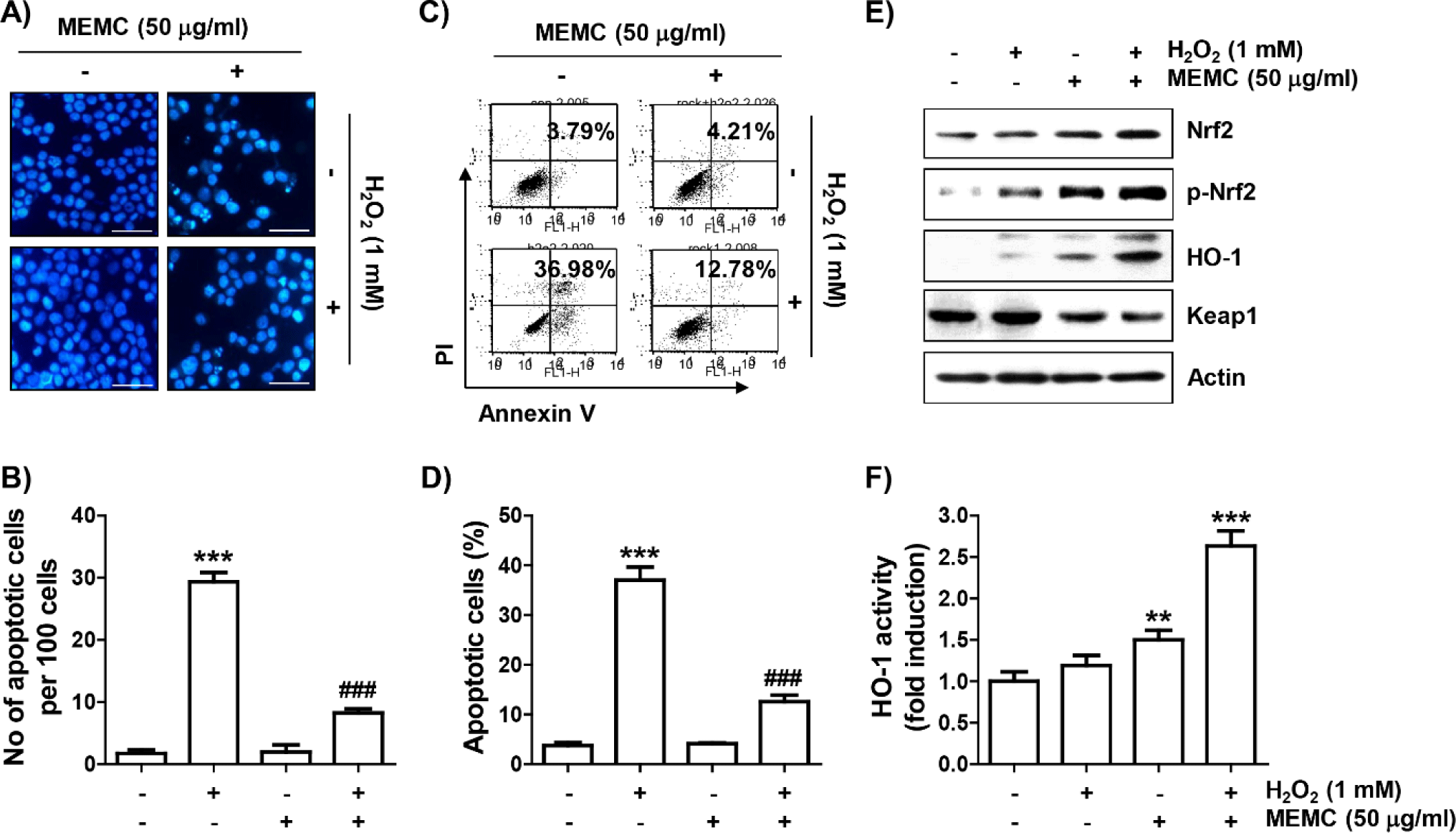
To confirm that MEMC prevented H2O2-induced apoptosis, nuclear morphology change observation by DAPI staining and flow cytometry analysis by annexin V/PI staining were performed, respectively. As presented in Fig. 5A and 5B, in H2O2-exposed C2C12 cells, morphological changes such as nuclear fragmentation and chromatin condensation, which are characteristically observed in apoptosis-induced nuclei, were clearly observed. However, MEMC pretreatment markedly attenuated these nuclear morphological changes. In parallel with this, flow cytometry results showed that much more apoptosis was induced in H2O2-treated cells than in control cells (Fig. 5C and 5D). However, the induction of apoptosis by H2O2 was greatly attenuated by MEMC pretreatment, similar to the reduction of sub-G1 cell population.
fas-26-1-35-g5
Fig. 5. MEMC protects cells against apoptosis and activates Nrf2 in H2O2-treated C2C12 cells. (A) After DAPI staining, images of representative nuclei are presented (× 400). (B and C) To measure the frequency of apoptosis, flow cytometry was performed after double staining with annexin V/PI. Representative histograms (B) and quantitative data (C) were shown. (D) After extracting cell lysate for each treatment group, expression levels of presented proteins were investigated through immunoblotting. (E) HO-1 activity is presented as a relative value. (B, D and F) The statistical significance evaluation results were presented (**p< 0.01 and ***p< 0.001 vs. control group; ###p< 0.001 vs. H2O2-treated cells). MEMC, methanol extract of Myelophycus caespitosus; H2O2, hydrogen peroxide; Nrf2, nuclear factor-erythroid-2 related factor 2; p-Nrf2, phosphorylation of Nrf2; DAPI, 4,6-diamidino-2-phenolindole; PI, propidium iodide; HO-1, heme oxygenase-1; Keap1, Kelch-like ECH-associated protein-1.
Download Original Figure
Nuclear factor-erythroid-2 related factor 2 (Nrf2)/heme oxygenase-1 (HO-1) signaling is further activated by methanol extract of Myelophycus caespitosus (MEMC) in hydrogen peroxide (H2O2)-treated cells

We subsequently investigated whether activation of the Nrf2/HO-1 axis was correlated with the antioxidant capacity of MEMC. As shown in Fig. 5E, the levels of Nrf2 as well as its phosphorylated form (p-Nrf2) were slightly increased by treatment with MEMC or H2O2 alone. However, their expression levels were remarkedly enhanced in H2O2-treated cells after MEMC pretreatment, whereas levels of Kelch-like ECH-associated protein-1 (Keap1) were much lower. Furthermore, the expression and activity of HO-1 were markedly promoted in cells treated with MEMC and H2O2 (Fig. 5E and 5F), suggesting that the Nrf2/HO-1 signaling pathway was further increased by MEMC in H2O2 -treated cells.

Activation of heme oxygenase-1 (HO-1) is involved in mitigating hydrogen peroxide (H2O2)-induced cytotoxicity by methanol extract of Myelophycus caespitosus (MEMC)

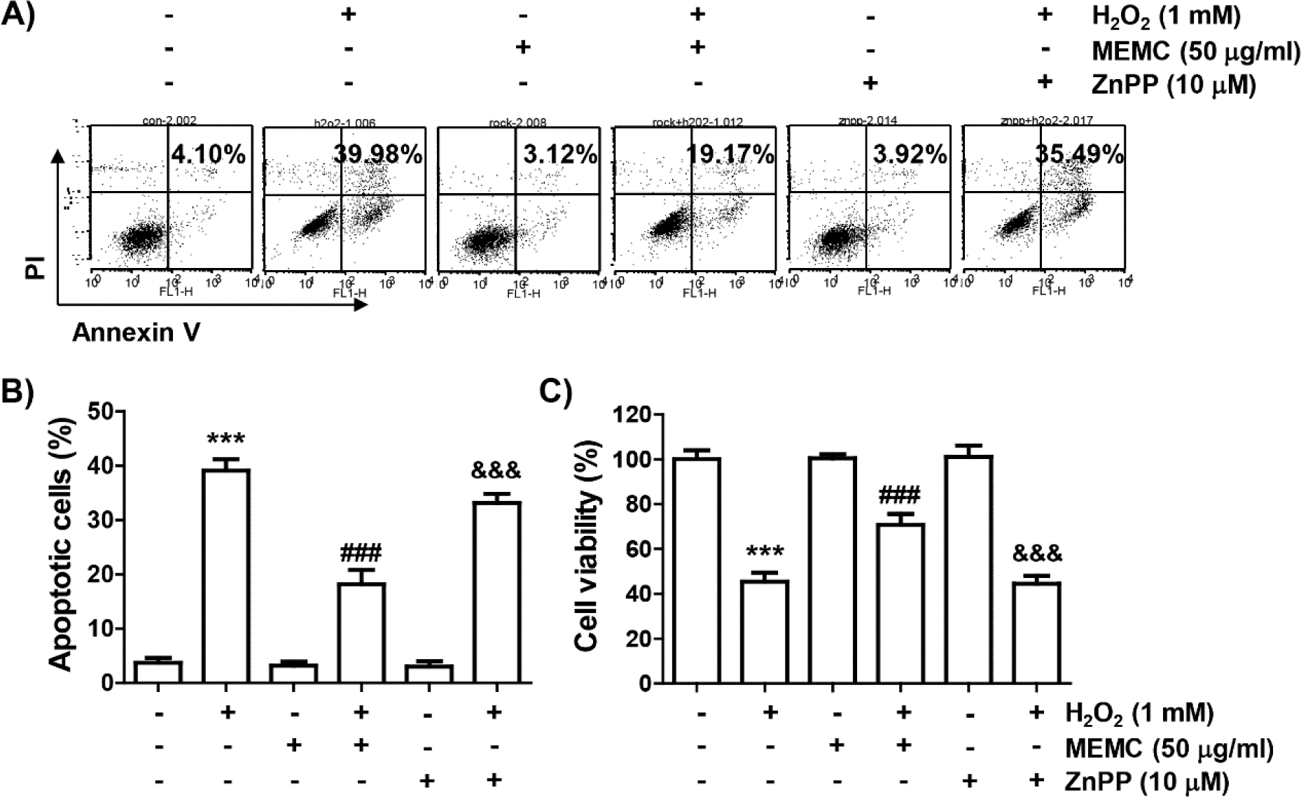
Finally, we used ZnPP, a selective inhibitor of HO-1, to evaluate whether activation of HO-1 by MEMC in H2O2-treated C2C12 cells was directly correlated with the cytotoxicity blocking effect of MEMC. As shown in Fig. 6A, 6B and 6C, the blocking effect of MEMC on apoptosis induced in cells exposed to H2O2 was markedly counteracted in the presence of ZnPP and pretreatment of ZnPP significantly abolished the anti-proliferative effect of MEMC in H2O2-stimulated cells. Consequently, these observations demonstrate that Nrf2-mediated activation of HO-1 at least served as an upstream signal to the inhibitory action of MEMC against H2O2-induced cytotoxicity of C2C12 cells.
fas-26-1-35-g6
Fig. 6. Effect of ZnPP, an HO-1 inhibitor, on the prevention of H2O2-induced cytotoxicity by MEMC in C2C12 cells. (A and B) To measure the frequency of apoptosis, flow cytometry was performed after double staining with annexin V/PI. Representative histograms (A) and quantitative results (B) were shown. (C) MTT assay was performed to assess cell viability. (B and C) The statistical significance evaluation results were presented (***p< 0.001 vs. control group; ###p< 0.001 vs. H2O2-treated cells; &&&p< 0.001 vs. with MEMC + H2O2 treatment group). MEMC, methanol extract of Myelophycus caespitosus; H2O2, hydrogen peroxide; ZnPP, zinc protoporphyrin IX; HO-1, heme oxygenase-1; PI, propidium iodide; MTT, 3-(4,5-dimethylthiazol-2-yl)-2,5-diphenyltetra-zolium bromide.
Download Original Figure

Discussion

In the current study, we induced oxidative stress using H2O2 to investigate whether MEMC could protect C2C12 myoblasts from oxidative injury and found that H2O2 treatment induced DNA damage, cell cycle arrest and apoptotic cell death through an increase in ROS generation. However, MEMC was found to be able to block H2O2-induced cytotoxicity while scavenging ROS as an Nrf2 activator.

Induction of cytotoxicity, including DNA damage and apoptosis, by oxidative stimuli is mostly accompanied by cell cycle arrest. Many previous studies have indicated that the cytotoxic effect of H2O2 is closely related to cell cycle arrest at the G2/M phase (Ding et al., 2019; Mubarok et al., 2022). Similar results were also observed in this study using C2C12 myoblasts due to an increase of Cdk inhibitor p21, a negative regulator of cell cycle progression, and decreases of cyclin A and cyclin B1 required for G2 to M phase progression (Dai et al., 2019; Taylor & Stark, 2001). However, H2O2-induced changes of these cell cycle regulators and cell cycle arrest were alleviated by MEMC pretreatment. In addition, the increase in the number of cells corresponding to the sub-G1 stage, which is used as an indicator of apoptosis, was significantly decreased in MEMC-pretreated cells. A similar trend was observed in NAC-pretreated cells. Therefore, it could be predicted that the protective effect of MEMC against H2O2-induced cytotoxicity was associated with inhibition of ROS generation. Similar to NAC pretreatment, MEMC pretreatment completely attenuated the ROS production by H2O2. Moreover, in H2O2-treated C2C12 cells, the formation of comet tails and p-γH2AX, as indicators of oxidative DNA damage (Kopp et al., 2019; Rahmanian et al., 2021), were effectively inhibited by MEMC. Therefore, our results suggest that the ROS scavenging ability of MEMC is responsible for H2O2-induced blockade of cytotoxicity in C2C12 cells.

Apoptosis is largely divided into extrinsic and intrinsic pathways. Overload of ROS by oxidative stress can depolarize mitochondrial membrane and contribute to the activation of the intrinsic apoptosis pathway (Bock & Tait, 2020; Tiwari et al., 2022; Urbani et al., 2021). This results in the loss of MMP, an indicative of mitochondrial dysfunction, and cytosolic release of cytochrome c. It is well known that cytochrome c can activate the caspase cascade required for the mitochondria-mediated intrinsic pathway, resulting in degradation of caspase-dependent proteins such as PARP, thereby terminating apoptosis (Sarwar et al., 2020; Tiwari et al., 2022). In this study, H2O2-induced reduction of MMP, cytosolic release of cytochrome c and cleavage of PARP were largely blocked by MEMC pretreatment. Furthermore, H2O2-induced changes in Bcl-2 family proteins were maintained at control levels after MEMC pretreatment. Accumulated prior studies have shown that the intrinsic pathway is critically controlled by proteins belonging to the Bcl-2 family. Among them, anti-apoptotic proteins including Bcl-2 are essential to maintain stability of the mitochondrial membrane barrier, whereas anti-apoptotic proteins such as Bax are key executors of mitochondrial poration, thereby enhancing mitochondrial membrane permeability and releasing mitochondrial cytochrome c (Bock & Tait, 2020; Tiwari et al., 2022). Several studies have demonstrated that the activation of mitochondria-mediated pathway observed in myoblasts exposed to oxidative stress is typically ROS-dependent (Choi, 2021; Drysch et al., 2021; Yu et al., 2019). In particular, apoptosis, which is observed relatively high in myogenic precursor cells derived from aged muscles, is the cause of weakening of skeletal muscle regeneration, which is related to a high level of ROS (Di Filippo et al., 2016; Fulle et al., 2013; Ji, 2015). These findings well support our data that MEMC can prevent C2C12 myoblasts from apoptosis via suppression of the intrinsic apoptotic pathway by blocking the generation of ROS caused by oxidative stress. Therefore, we propose that treatment with MEMC might contribute to the maintenance of myoblast function and their differentiation into myocytes.

Nrf2 is a redox-sensitive transcription factor that can enhance antioxidant capacity by regulating the expression of phase II detoxification enzymes (Jenkins & Gouge, 2021; Tonelli et al., 2018). When cells are treated with Nrf2 activators or exposed to oxidative stress, Nrf2 must be phosphorylated for nuclear translocation after dissociation from Keap1, a negative regulator of Nrf2, to promote transcriptional activity of Nrf2-dependent genes. As a representative downstream factor of Nrf2, HO-1 can decompose heme into biliverdin, carbon monoxide and free iron. The produced biliverdin is then converted to bilirubin, which has an antioxidant activity (Jenkins & Gouge, 2021; Yu et al., 2018). These findings indicate that discovering substances that can activate the Nrf2/HO-1 axis might be one of the appropriate strategies to counteract cellular damage caused by oxidative stress. Recently, numerous studies have suggested that various natural products have antioxidant properties that can inhibit oxidative damage-mediated apoptosis in myoblasts through activation of the Nrf2/HO-1 signaling (Rui et al., 2022; Wu et al., 2022; Xu et al., 2021; Zhang et al., 2021). Therefore, we investigated whether MEMC could activate Nrf2 and found that the level of p-Nrf2 expression indicating that Nrf2 was activated was clearly upregulated by H2O2 in MEMC-pretreated C2C12 cells. In addition, in MEMC-treated C2C12 cells under oxidative conditions, the activity and the expression of HO-1 was significantly enhanced, while Keap1 expression was decreased. However, the apoptosis blocking and cytoprotective potential by MEMC in H2O2-treated cells was significantly counteracted by ZnPP, an HO-1 inhibitor, suggesting that activation of HO-1 was at least responsible for the blockade of H2O2-induced oxidative damage by MEMC. Our results support previous studies showing that antioxidant activities of various seaweed extracts and their constituents are due to activation of the Nrf2/HO-1 signaling (Jayawardena et al., 2018; Kang et al., 2015; Lekshmi et al., 2019; Niu et al., 2020; Park et al., 2018). Our results are also in good agreement with those of previous studies showing that Nrf2-mediated activation of HO-1 in myoblasts can act as a protective mechanism against apoptosis induced after mitochondrial dysfunction caused by oxidative stimulation (Li et al., 2020; Park et al., 2021; Song et al., 2022).

Taken together, the present results showed that MEMC could alleviate cell cycle arrest, DNA damage, and apoptosis by mitigating H2O2-induced mitochondrial damage as an ROS scavenger in C2C12 myoblasts. In addition, MEMC as an activator of Nrf2 might contribute to the blockade of oxidative damage through activation of HO-1, indicating that MEMC has a high potential application in the maintenance of myoblast function against oxidative damage. However, further studies are required to determine upstream signaling pathways regulating the activity of Nrf2 by MEMC and other antioxidant signals that might be involved in its antioxidant activity.

Competing interests

No potential conflict of interest relevant to this article was reported.

Funding sources

This research was supported by Korea Institute of Marine Science & Technology Promotion (KIMST) funded by the Ministry of Oceans and Fisheries, Korea (20220488).

Acknowledgements

Not applicable.

Availability of data and materials

Upon reasonable request, the datasets of this study can be available from the corresponding author.

Ethics approval and consent to participate

This article does not require IRB/IACUC approval because there are no human and animal participants.

References

1.

Abrigo J, Simon F, Cabrera D, Vilos C, Cabello-Verrugio C. Mitochondrial dysfunction in skeletal muscle pathologies. Curr Protein Pept Sci. 2019; 20:536-46

2.

Besednova NN, Andryukov BG, Zaporozhets TS, Kuznetsova TA, Kryzhanovsky SP, Ermakova SP, et al. Molecular targets of brown algae phlorotannins for the therapy of inflammatory processes of various origins. Mar Drugs. 2022; 20:243

3.

Bock FJ, Tait SWG. Mitochondria as multifaceted regulators of cell death. Nat Rev Mol Cell Biol. 2020; 21:85-100

4.

Carroll AR, Copp BR, Davis RA, Keyzers RA, Prinsep MR. Marine natural products. Nat Prod Rep. 2021; 38:362-413

5.

Choi YH. Trans-cinnamaldehyde protects C2C12 myoblasts from DNA damage, mitochondrial dysfunction and apoptosis caused by oxidative stress through inhibiting ROS production. Genes Genomics. 2021; 43:303-12

6.

Choi YH. Tacrolimus induces apoptosis in leukemia Jurkat cells through inactivation of the reactive oxygen species-dependent phosphoinositide-3-kinase/Akt signaling pathway. Biotechnol Bioprocess Eng. 2022; 27:183-92

7.

Dai Y, Jin F, Wu W, Kumar SK. Cell cycle regulation and hematologic malignancies. Blood Sci. 2019; 1:34-43

8.

Di Filippo ES, Mancinelli R, Pietrangelo T, La Rovere RM, Quattrocelli M, Sampaolesi M, et al. Myomir dysregulation and reactive oxygen species in aged human satellite cells. Biochem Biophys Res Commun. 2016; 473:462-70

9.

Di Meo S, Iossa S, Venditti P. Skeletal muscle insulin resistance: role of mitochondria and other ROS sources. J Endocrinol. 2017; 233:R15-42

10.

Ding Y, Zhang Z, Yue Z, Ding L, Zhou Y, Huang Z, et al. Rosmarinic acid ameliorates H2O2-induced oxidative stress in L02 cells through MAPK and Nrf2 pathways. Rejuvenation Res. 2019; 22:289-98

11.

Drysch M, Schmidt SV, Becerikli M, Reinkemeier F, Dittfeld S, Wagner JM, et al. Myostatin deficiency protects C2C12 cells from oxidative stress by inhibiting intrinsic activation of apoptosis. Cells. 2021; 10:1680

12.

Fulle S, Sancilio S, Mancinelli R, Gatta V, Di Pietro R. Dual role of the caspase enzymes in satellite cells from aged and young subjects. Cell Death Dis. 2013; 4:e955

13.

Hanyuda T, Aoki S, Kawai H. Reinstatement of Myelophycus caespitosus (Scytosiphonaceae, Phaeophyceae) from Japan. Phycol Res. 2020; 68:126-34

14.

Jayasooriya RGPT, Kang CH, Jang YJ, Kang SH, Dilshara MG, Choi YH, et al. Methanol extract of Myelophycus caespitosus inhibits the inflammatory response in lipopolysaccharide-stimulated BV2 microglial cells by downregulating NF-kB via Inhibition of the Akt signaling pathway. Trop J Pharm Res. 2012; 11:917-24

15.

Jayawardena TU, Asanka Sanjeewa KK, Shanura Fernando IP, Ryu BM, Kang MC, Jee Y, et al. Sargassum horneri (Turner) C. Agardh ethanol extract inhibits the fine dust inflammation response via activating Nrf2/HO-1 signaling in RAW 264.7 cells. BMC Complement Altern Med. 2018; 18:249

16.

Jenkins T, Gouge J. Nrf2 in cancer, detoxifying enzymes and cell death programs. Antioxidants. 2021; 10:1030

17.

Jeong MJ, Lim DS, Kim SO, Park C, Leem SH, Lee H, et al. Protection of oxidative stress-induced DNA damage and apoptosis by rosmarinic acid in murine myoblast C2C12 cells. Biotechnol Bioprocess Eng. 2022; 27:171-82

18.

Ji LL. Redox signaling in skeletal muscle: role of aging and exercise. Adv Physiol Educ. 2015; 39:352-9

19.

Kang JS, Choi IW, Han MH, Lee DS, Kim GY, Hwang HJ, et al. The cytoprotective effect of Petalonia binghamiae methanol extract against oxidative stress in C2C12 myoblasts: mediation by upregulation of heme oxygenase-1 and nuclear factor-erythroid 2 related factor 2. Mar Drugs. 2015; 13:2666-79

20.

Kopp B, Khoury L, Audebert M. Validation of the γH2AX biomarker for genotoxicity assessment: a review. Arch Toxicol. 2019; 93:2103-14

21.

Lekshmi VS, Rauf AA, Muraleedhara Kurup G. Sulfated polysaccharides from the edible marine algae Padina tetrastromatica attenuates isoproterenol-induced oxidative damage via activation of PI3K/Akt/Nrf2 signaling pathway: an in vitro and in vivo approach. Chem Biol Interact. 2019; 308:258-68

22.

Li J, Yang Q, Han L, Pan C, Lei C, Chen H, et al. C2C12 mouse myoblasts damage induced by oxidative stress is alleviated by the antioxidant capacity of the active substance phloretin. Front Cell Dev Biol. 2020; 8:541260

23.

Liu Y, Guo Z, Wang S, Liu Y, Wei Y. Fucoxanthin pretreatment ameliorates visible light-induced phagocytosis disruption of RPE cells under a lipid-rich environment via the Nrf2 pathway. Mar Drugs. 2021; 20:15

24.

Mubarok W, Elvitigala KCML, Nakahata M, Kojima M, Sakai S. Modulation of cell-cycle progression by hydrogen peroxide-mediated cross-linking and degradation of cell-adhesive hydrogels. Cells. 2022; 11:881

25.

Mukherjee S, Park JP, Yun JW. Carboxylesterase3 (Ces3) interacts with bone morphogenetic protein 11 and promotes differentiation of osteoblasts via Smad1/5/9 pathway. Biotechnol Bioprocess Eng. 2022; 27:1-16

26.

Niu T, Fu G, Zhou J, Han H, Chen J, Wu W, et al. Floridoside exhibits antioxidant properties by activating HO-1 expression via p38/ERK MAPK pathway. Mar Drugs. 2020; 18:105

27.

Park C, Hong SH, Shin SS, Lee DS, Han MH, Cha HJ, et al. Activation of the Nrf2/HO-1 signaling pathway contributes to the protective effects of Sargassum serratifolium extract against oxidative stress-induced DNA damage and apoptosis in SW1353 human chondrocytes. Int J Environ Res Public Health. 2018; 15:1173

28.

Park C, Lee H, Hong S, Molagoda IMN, Jeong JW, Jin CY, et al. Inhibition of lipopolysaccharide-induced inflammatory and oxidative responses by trans-cinnamaldehyde in C2C12 myoblasts. Int J Med Sci. 2021; 18:2480-92

29.

Pradhan B, Nayak R, Patra S, Jit BP, Ragusa A, Jena M. Bioactive metabolites from marine algae as potent pharmacophores against oxidative stress-associated human diseases: a comprehensive review. Molecules. 2020; 26:37

30.

Rahmanian N, Shokrzadeh M, Eskandani M. Recent advances in γH2AX biomarker-based genotoxicity assays: a marker of DNA damage and repair. DNA Repair (Amst). 2021; 108:103243

31.

Rui Y, Li S, Luan F, Li D, Liu R, Zeng N. Several alkaloids in Chinese herbal medicine exert protection in acute kidney injury: focus on mechanism and target analysis. Oxid Med Cell Longev. 2022; 2022:2427802

32.

Sambasivan R, Tajbakhsh S. Adult skeletal muscle stem cells. Results Probl Cell Differ. 2015; 56:191-213

33.

Santa-Gonzalez GA, Gomez-Molina A, Arcos-Burgos M, Meyer JN, Camargo M. Distinctive adaptive response to repeated exposure to hydrogen peroxide associated with upregulation of DNA repair genes and cell cycle arrest. Redox Biol. 2016; 9:124-33

34.

Sarwar MS, Xia YX, Liang ZM, Tsang SW, Zhang HJ. Mechanistic pathways and molecular targets of plant-derived anticancer ent-kaurane diterpenes. Biomolecules. 2020; 10:144

35.

Sivandzade F, Bhalerao A, Cucullo L. Analysis of the mitochondrial membrane potential using the cationic JC-1 dye as a sensitive fluorescent probe. Bio Protoc. 2019; 9:e3128

36.

Song BR, Alam MB, Lee SH. Terpenoid-rich extract of Dillenia indica L. bark displays antidiabetic action in insulin-resistant C2C12 cells and STZ-induced diabetic mice by attenuation of oxidative stress. Antioxidants (Basel). 2022; 11:1227

37.

Steinbacher P, Eckl P. Impact of oxidative stress on exercising skeletal muscle. Biomolecules. 2015; 5:356-77

38.

Taylor WR, Stark GR. Regulation of the G2/M transition by p53. Oncogene. 2001; 20:1803-15

39.

Tiwari S, Dewry RK, Srivastava R, Nath S, Mohanty TK. Targeted antioxidant delivery modulates mitochondrial functions, ameliorates oxidative stress and preserve sperm quality during cryopreservation. Theriogenology. 2022; 179:22-31

40.

Tonelli C, Chio IIC, Tuveson DA. Transcriptional regulation by Nrf2. Antioxid Redox Signal. 2018; 29:1727-45

41.

Urbani A, Prosdocimi E, Carrer A, Checchetto V, Szabò I. Mitochondrial ion channels of the inner membrane and their regulation in cell death signaling. Front Cell Dev Biol. 2021; 8:620081

42.

Wu AG, Yong YY, Pan YR, Zhang L, Wu JM, Zhang Y, et al. Targeting Nrf2-mediated oxidative stress response in traumatic brain injury: therapeutic perspectives of phytochemicals. Oxid Med Cell Longev. 2022; 2022:1015791

43.

Xu W, Zhu H, Hu B, Cheng Y, Guo Y, Yao W, et al. Echinacea in hepatopathy: a review of its phytochemistry, pharmacology, and safety. Phytomedicine. 2021; 87:153572

44.

Yu LM, Zhang WH, Han XX, Li YY, Lu Y, Pan J, et al. Hypoxia-induced ROS contribute to myoblast pyroptosis during obstructive sleep apnea via the NF-κB/HIF-1α signaling pathway. Oxid Med Cell Longev. 2019; 2019:4596368

45.

Yu Y, Cui Y, Niedernhofer LJ, Wang Y. Occurrence, biological consequences, and human health relevance of oxidative stress-induced DNA damage. Chem Res Toxicol. 2016; 29:2008-39

46.

Yu ZY, Ma D, He ZC, Liu P, Huang J, Fang Q, et al. Heme oxygenase-1 protects bone marrow mesenchymal stem cells from iron overload through decreasing reactive oxygen species and promoting IL-10 generation. Exp Cell Res. 2018; 362:28-42

47.

Zhang Q, Liu J, Duan H, Li R, Peng W, Wu C. Activation of Nrf2/HO-1 signaling: an important molecular mechanism of herbal medicine in the treatment of atherosclerosis via the protection of vascular endothelial cells from oxidative stress. J Adv Res. 2021; 34:43-63