RESEARCH ARTICLE

Morphological variations of Gnathonemus petersii (Günther, 1862) (Pisces, Mormyridae) based on museum preserved adult specimens

Alamrew Eyayu Zeleke 1 , * https://orcid.org/0000-0003-1029-5191
Author Information & Copyright
1Department of Biology, Debre Tabor University, Debre Tabor 272, Ethiopia
*Corresponding author: Alamrew Eyayu Zeleke, Department of Biology, Debre Tabor University, Debre Tabor 272, Ethiopia, Tel: +251-927714676, E-mail:eyayualam2010@gmail.com

Copyright © 2025 The Korean Society of Fisheries and Aquatic Science. This is an Open Access article distributed under the terms of the Creative Commons Attribution Non-Commercial License (http://creativecommons.org/licenses/by-nc/4.0/) which permits unrestricted non-commercial use, distribution, and reproduction in any medium, provided the original work is properly cited.

Received: Oct 17, 2024; Revised: Dec 04, 2024; Accepted: Dec 30, 2024

Published Online: Jun 30, 2025

Abstract

Examination of the morphometric (measurable) and meristic (countable) characteristics in fish is important for the differentiation of taxonomic units to spot differences in fishes. The taxonomical study of African freshwater mormyrids is of great interest because of their morphological diversification. Therefore, the study of variations in morphological traits among fish species has been conventionally based on comparisons of adults. In this study, morphological variations were studied for Gnathonemus petersii based on museum deposited adult specimens collected from African rivers. Specimens were previously (2015) collected, identified, tagged, and deposited in the Royal Museum for Central Africa. The study was based on 29 measurable and eight countable traits along with character definitions to compare morphological variations in G. petersii. Accordingly, the 29 measurable traits were measured using a caliper, and meristic enumerations were supported by dissecting microscope. Overall, a total of 119 individuals of G. petersi were measured and enumerated for their morphometrics and meristics, respectively, to interpret intraspecific variations. Counts of pelvic and pectoral fins for all specimens were the same (6 and 10, respectively) and excluded for the analysis. However, specimens collected from the Wouri and Sanaga Rivers showed some grouping being isolated from Nigerian specimens. Out of the 29 morphometric variables measured, 18 variables showed slight differences. In the present study, the test for clear separation analysis applied for the G. petersi species using morphometric measurements showed slight separations in between specimens collected from Congo and Sanaga. Generally, the morphometric measurements, which represent standard length (%SL) and head length (%HL) were the most discriminate characters used in the present analysis. Although systematic ichthyologists continue to depend heavily on morphological variations for taxonomic characters, the value and availability of genetic, physiological, behavioral, and ecological data need to be used in modern fish nomenclature.

Keywords: Morphology; Museum; Taxonomy; Traits; Variation

Introduction

The family Mormyridae belongs to the most primitive group of teleostean fishes- the Osteoglossomorpha (Nelson et al., 2016). Fishes of this family are endemic to African freshwater and include 22 genera and 230 species (Eschmeyer et al., 2020; Froese & Pauly, 2019; Moyle & Cech, 2000). In terms of genus and species diversity, the family Mormyridae exceeds all other extant Osteoglossomorph lineages (Simanovsky et al., 2020). All fish in the family produce weak electric discharges that can sense perturbations and be used as a means of communication (Carlson & Arnegard, 2011; Engelmann et al., 2009).

Mormyrids are known for a few notable structural and physiological characteristics, with the possession of a narrow caudal peduncle and a deeply forked caudal fin (Bell, 1989). They are diverse in morphology and unique in some external structures. The possession of mouths of highly variable form and often trunks like in some genera (e.g., Gnathonemus) is common. Phylogenetic trends are difficult to trace in mormyrids (Bass, 1986). Thus, morphological and molecular techniques have aided in further phylogenetic analysis (Lavoué et al., 2003). Molecular studies have provided a well-supported tree for the major mormyrid lineages (Alves-Gomes & Hopkins, 1997; Lavoué et al., 2003; Sullivan et al., 2002). The morphological works have also been employed in resolving fish taxonomical illusions (e.g., Abdurahman et al., 2016; Gelsano & Demayo, 2022; Kerschbaumer & Sturmbauer, 2011; Strauss & Bond, 1990). Both the molecular and morphological analysis in mormyridae taxonomical studies agree on: (1) mormyrid monophyly, (2) sister-group relationship between mormyridae and gymnarchidae, and (3) the basal division of the family mormyridae into two subfamilies (Mormyrinae and Petrocephalinae; Lavoué et al., 2003).

Among others, the ‘elephant-nose fish’ Gnathonemus petersii (Günther, 1862) is an African freshwater mormyrid and is native to the rivers of west and central Africa, in particular the river basins of lower Niger, Ogun and upper Chari. The species has a vast distribution and has been recorded in Mali, Benin, Niger, Nigeria, Chad, the Central African Republic, Cameroon, Democratic Republic of the Congo, and Zambia. In Lower Guinea, the species are found in the Cross, Mungo, Wouri, Lokoundjé, and Lower Sanaga Rivers (Nelson et al., 2016). The species is widely distributed throughout central Africa, from the Niger Delta to the Congo River basin. Thus, a wide distribution of the species suggests re-visionary work may uncover multiple species (Nelson et al., 2016).

G. petersii prefers slowly moving rivers and pools with muddy bottoms covered with submerged macrophytes (Engelmann et al., 2009). The species is a dark brown to black in color, laterally compressed (averaging 23–25 cm, total length [TL]), with a rear dorsal fin and anal fin of the same length with a forked caudal fin (Fig. 1). The species has two stripes on its lower pendicular and a trunk-like protrusion on the head. This protrusion is not a nose, but rather a sensitive extension of the mouth used for self-defense, communication, navigation, and finding prey. G. petersii can be recognized easily by the black brown coloration of two distinct bands in the shape of parentheses “()” running from the origins of dorsal to anal fins (Fig. 1). The species feeds on small worms and aquatic invertebrates such as mosquito larvae in the natural habitat (Nwani et al., 2011). The species has good low light vision; the eyes use a combination of photonic crystals, parabolic mirrors, and a clustered arrangement of rods and cones (Engelmann et al., 2009). As a unique feature in mormyrids, G. petersii possess a weak electric field from its electroreceptors (Engelmann et al., 2009).

fas-28-6-372-g1
Fig. 1. Gnathonemus petersii (Günther, 1862)- showing peculiar color patterns and mouth protrusion (left) and preserved specimen during measurement (right).
Download Original Figure

The use of morphological parameters in taxonomical studies is common; this technique is very cost-effective and could be promoted as a tool for aiding in species identification, particularly for morpologically diverse fish species. Information on the external morphology of fishes is used in many taxonomical works (Hubbs & Lagler, 1958; Lagler et al., 1977; Miller & Lea, 1972; Moyle & Cech, 2000; Strauss & Bond, 1990; Trautman 1981). The morphology of fish is a result of adaptations to several forces. Environmental influences cause variations in the general structure, and thus measuring morphological variations in fish is crucial to resolving the question of taxonomical uncertainties (Strauss & Bond, 1990). The morphometric and countable traits of fish historically have been used for taxonomic and evolutionary studies (Brosse et al., 2021). Despite the value and availability of genetic, physiological, behavioral, and ecological data for such studies, systematic ichthyologists continue to depend heavily on morphology for taxonomic characters (Abdurahman et al., 2016; Gelsano & Demayo, 2022; Strauss & Bond, 1990). In addition to measurable and countable traits, species have characteristic shapes, sizes, pigmentation patterns, disposition of fins, and other external features that aid in recognition, identification, and classification (Gelsano & Demayo, 2022). Therefore, the present study aimed to characterize morphological variations in G. petersii collected from different localities in western and central Africa based on morphometric measurements, meristic counts, and observations on color pattern, positions of mouth, and fin arrangements.

Materials and Methods

Study specimens

A total of 119 fish specimens used in this study were obtained from museum preserved G. petersii deposited in the Royal Museum for Central Africa (RMCA; Ichthyology section). The specimens were collected from river basins of western Africa (Cameroon, DR Congo, Nigeria) and the Central Africa Republic in 2015. Each specimen was identified to species level and soaked in alcohol solutions. Damaged specimens due to preservation procedures were excluded during the analysis.

Character definition, measurements, and counting protocols

Museum collections of undamaged G. petersii voucher specimens from the RMCA were used for this study. Regardless of the method used to acquire morphometric measurements, care was taken to ensure that specimens were not abnormal or otherwise deformed in shape or posture because of preservation. Linear morphometric measurements were carried out from the left lateral side of the fish. Over all, 119 unbroken specimens previously identified as G. petersii were measured and analyzed for morphological variations (Table 1). About 29 important morphometric, eight meristic, and four descriptive characters were analyzed during the study (Table 1). Character definitions, methods for making counts, and measurements were based on Boden et al. (1997). The morphometric measurements were taken related to general body features, length, mouth, chin lobe, and head related regions (Strauss & Bond, 1990), whereas meristic parameters include fin ray and scale counts (Abdurahman et al., 2016). A dial calliper diameter with a precision level of 0.01 mm was used for morphometric measurements. Meristic counts were taken with the help of hand lenses and microscopes. The morphometric characters measured and their respective definitions are presented in Table 1.

Table 1. Morphometrics, meristics, and descriptive morphological characters of Gnathonemus petersii used for the present study
Characters considered Character definition
Morphometrics
 Standard length (SL) Distance from tip of snout to base of caudal fin
 Body depth (BD) Vertical depth of body taken from the anterior base of the pelvic fin (maximum point)
 Head length (HL) Distance from snout tip to posterior end of opercula
 Head depth (HD) Vertical height at anterior end of gill cover
 Head width (HW) Distance between posterior ends of the two opercula
 Snout length (SnL) From anterior tip of snout to end of anterior eye border
 Eye diameter (ED) Circumference of the eye
 Inter orbital width (IoW) Width between the eyes
 Post orbital length (PoL) Distance from posterior border of the eye to posterior end of operculum
 Distance between nostrils (DNN) Distance between anterior and posterior nostrils
 Distance between nostril and eye (DNE) Distance between posterior nostril and eye
 Mouth width (MW) Width between upper and lower jaws
 Chin lobe length (ClL) Length of the lobe extension from lower jaw
 Predorsal distance (PDD) From anterior snout tip to dorsal fin anterior base
 Preanl distance (PAD) From anterior tip of snout to anal fin anterior base
 Prepelvic distance (PPlD) From anterior snout tip to anterior base of pelvic fin
 Prepectoral distance (PPcD) From tip of snout to anterior base of pectoral fin
 Dorsal fin length (DFL) Anterior to posterior ends of the fin
 Dorsal fin height (DFH) Longest ray length
 Anal fin length (AFL) Anterior to posterior ends of the fin
 Anal fin height (AFH) Longest ray length
 Pelvic fin length (PlFL) Anterior to posterior ends of the fin
 Pectoral fin length (PcFL) Anterior to posterior ends of the fin
 Distance between pelvic and anal fin (D-PlF-AF) Distance between anterior bases of the fins
 Distance between pectoral and anal fin (D-PcF-AF) Distance between anterior bases the two fins
 Distance between pectoral and pelvic fin (D-PcF-PlF) Distance between anterior bases the two fins
 Distance between dorsal and anal fin (D-DF-AF) Distance between anterior bases the two fins
 Caudal peduncle length (CPL) From posterior end of the anal fine to insertion of the caudal fin
 Caudal peduncle height (CPH) Distance between posterior end to end of dorsal and anal fin
 Caudal peduncle depth (CPD) A minimum vertical distance of the caudal peduncle
Meristics
 Branched soft rays Counting all branched soft rays
 Anal fin rays (AFR) Counting all branched soft rays
 Pelvic fin rays (PvFR) Counting all branched soft rays
 Pectoral fin rays (PFR) Counting all branched soft rays
 Scales in the Lateral Line (SLL) Counting all scales along the lateral line
 Scales in the caudal peduncle (SCP) Scales counted around the CP from the narrowest part
 Scales between dorsal fin and lateral line (SDLL) Scales from lateral line to base of the anterior dorsal fin
 Scales between anal fin and lateral line (SALL) Scales from lateral line to base of the anterior anal fin
 Descriptive characters
Colour pattern Observation
 Mouth position Observation
 Position of nostrils Observation
 Fins arrangement (advancements) Observation
Download Excel Table
Methods of data analysis

Descriptive statistics were employed to ascertain the measured morphometric variables into a proportion of percentage in standard length (%SL) and head length (%HL). The principal component analysis (PCA), which is a widely used technique for the assessment of patterns of variations among a set of characters, was employed to examine variations in log transformed morphometric measurements. For the morphometric characters, a PCA analysis was used on percentage data in relation to %SL and %HL. Raw values of the meristic counts were used for the PCA analysis without conversions of proportions in to %SL and %HL. The PCA also permits the examination of size and shape differences independently and does not rely on ratios of measurements (Boden et al., 1997). Thus, the PCA was used to analyze and explore the data set in PAST software version 3.40 for this study.

Results and Discussion

Analysis of Meristic characteristics

During the present study, eight meristic characters were enumerated (Table 2). The variation among the counts in each parameters were rarely observed. The counts of pelvic and pectoral fins for all specimens were the same (6 and 10, respectively) and thus excluded for the analysis as recommended by Abdurahman et al. (2016). Therefore, this study was intended to trace the differences among the geographical locations where the specimens were collected. This is because meristic characters can be influenced substantially by environmental factors, especially temperature during early development. In the present study, however, as it is presented in Fig. 2, there was no clear separation on counts from different localities. However, some specimens collected from the Wouri and Sanaga Rivers showed some groupings being overlapped with some Nigerian and Congo specimens and tending to be aligned on the right side (Fig. 2). This might be attributed to the higher number of lateral line scales (LLS) in Wouri and Sanaga specimens over others, as reflected on first axis (PCA1) and second axis (PCA2) as a loading factor (Table 2). The proportion of the number of LLS was relatively higher in Sanaga specimens, followed by Wouri and Congo. By considering LLS, the study tried to encounter a pairwise comparison in each locality by categorizing specimens with less than 63 LLS as one group and more on the other group. However, clear separation was not observed on the number of LLS. The number of scales around the caudal peduncle was the same (eight) except for four specimens from Congo that had 10. Concerning the scales around the caudal peduncle, this study has tried to sort out a difference that had eight and 10 scales. However, there was no clear mark of isolation in between those having 8 and 10 (Fig. 2).
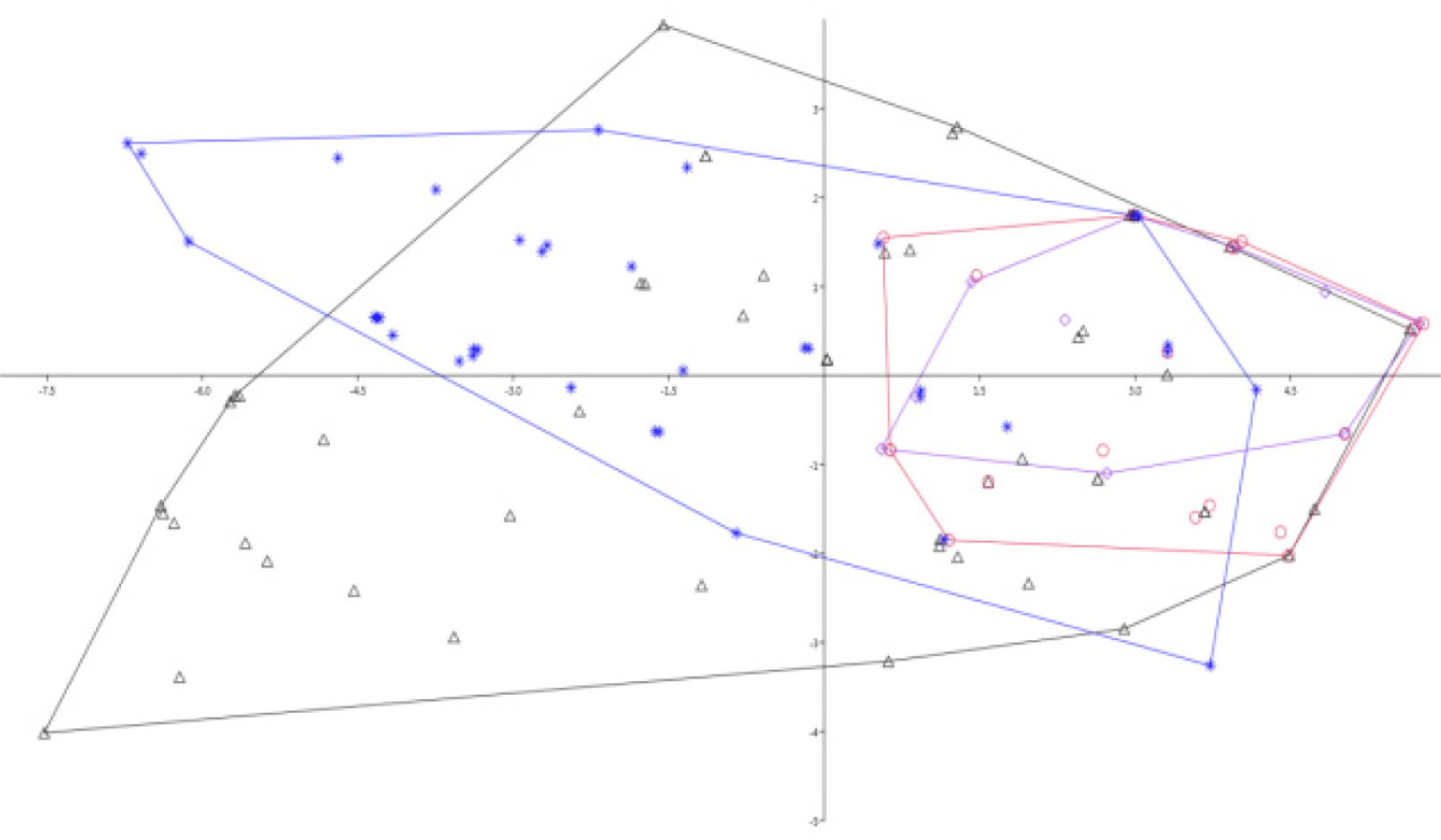
Table 2. Loading factors for PCA 1 and PCA 2 of a PCA on six meristics (n = 119)
Characters Loading factors
PCA1 PCA2 PCA3 PCA4
DFR 0.27059* 0.68337* –0.67298 0.03965
AFR 0.33437* 0.58671* 0.72203 –0.14043
SLL 0.9005* –0.42703* –0.07498 –0.028548
SCP 0.010118 0.039277 0.031586 –0.03238
SDLL 0.046965* 0.069617* 0.052944 0.69066
SALL 0.042185 –0.005246 0.12786 0.70699

* The most important loadings.

PCA, principal component analysis; PCA1, first axis; PCA2, second axis; PCA3, third axis; PCA4, fourth axis; AFR, anal fin rays; SLL, scales in the lateral line; SCP, scales in the caudal peduncle; SDLL, scales between dorsal fin and lateral line; SALL, scales between anal fin and lateral line.

Download Excel Table
fas-28-6-372-g2
Fig. 2. Plot of scores on the first and second axes of a PCA (PCA1- x axis and PCA2- y axis) on six raw merstics for all examined specimens (Triangle- Congo, star- Nigeria, circle- Wouri and diamond- Sanaga) (n = 119). PCA, principal component analysis; PCA1, first axis; PCA2, second axis.
Download Original Figure

Meristic characters are the body segments and other features, primarily fin rays and scales, that once, in evolutionary history, corresponded to the body segmentation (Strauss & Bond, 1990). Meristic is a quantitative enumeration of the characteristics (body parts) of fish, for example, the number of fins. Meristic characters that are counted as many as eight characters, among others: DFR, anal fin rays (AFR), pelvic fin rays (PvFR), pectoral fin rays (PFR), scales in the lateral line (SLL), scales in the caudal peduncle (SCP), scales between dorsal fin and lateral line (SDLL), and scales between anal fin and lateral line (SALL; Tables 1 and 2). Similar counting patterns and enumeration techniques were followed by Haryono (2000). Because the evaluation of meristic and other countable characters can be subjective, published accounts should explicitly define the criteria used in making such counts accordingly Hubbs & Lagler (1958). The PCA loading factors showed that the PCA1 and PCA2 explained more of the characters counted for this study and included DFR, AFR, SLL, and SDLL (Table 2). Meristic characters can be influenced substantially by environmental factors, especially by temperature during early development (Gelasno & Demayo, 2022). As a result, countable characters vary within and among species, so they are useful in describing or identifying fishes.

Analysis of morphometric measurements

Morphometric characters is a quantitative description, which have been successfully used for taxonomic inferences. Thus, documentation of morphological information is important to validate the taxonomical status and kinship relationship within or between species. A number of studies have shown that morphometric characters are suitable parameters for describing morphological variations among populations (Abdurahhman et al. 2016; Ahirwal et al., 2023; Biswal et al., 2018; Costa et al., 2003; Jacquemin & Pyron, 2016; Murta, 2000) and determining possible differences between individual unit stocks of the same species (Table 3; King, 2007). Out of the 29 morphometric variables measured for 119 samples, 18 variables showed significant differences. Without denying the role of genetic divergence, these differences might be caused by the differences in environment, as the studied specimens were sampled from different agroclimatic zones. Abdurahman et al. (2016) reported that freshwater fish display a range of morphological variations across a wide variety of physiological states and environmental conditions as a result of phenotypic plasticity. In the present study, the PCA analysis of measurements revealed that there was no clear separation among specimens collected from different localities. Morphometric variations are widely used to compare and differentiate among species and groups based on overall body type or distinctive anatomical forms in relation to one or two major body parts (Biswal et al., 2018; Jacquemin & Pyron, 2016).

Table 3. Mean and standard deviations (SD) of morphometric measurements in percentage
Congo Nigeria Wouri Sanaga
Mean ± SD Range Mean ± SD Range Mean ± SD Range Mean ± SD Range
SL 111.89 ± 42.52 55.24–246.34 120.61 ± 30.49 57.5–170.64 125.07 ± 29.38 86.23–186.72 154.44 ± 43.08 90.84–203.66
BD %SL 21.38 ± 2.26 17.72–26.16 23.92 ± 2.26 17.29–27.52 20.94 ± 2.05 16.91–25.03 21.49 ± 2.00 18.89 ± 25.92
HL %SL 25.98 ± 1.53 23.30–29.18 26.20 ± 1.00 23.55–27.75 24.83 ± 1.29 22.75–28.43 24.01 ± 0.55 23.44 ± 25.19
HD%HL 73.27 ± 6.51 61.35–87.54 80.67 ± 7.39 68.28–93.38 79.41 ± 7.19 68.52–96.15 84.87 ± 6.16 76.61–96.79
HW%HL 32.85 ± 3.99 27.79–45.79 33.94 ± 3.72 26.15–39.97 33.69 ± 2.91 26.81–39.03 34.77 ± 4.91 27.27–42.44
SnL %HL 34.12 ± 2.56 28.46–43.39 33.93 ± 2.06 29.15–38.99 35.47 ± 2.52 31.58–39.84 34.59 ± 2.66 29.90–38.83
ED %HL 16.19 ± 2.37 12.06–26.19 14.18 ± 1.57 11.86–19.19 15.99 ± 1.34 13.83–18.94 13.97 ± 1.58 12.13–16.87
IoW %HL 26.54 ± 2.51 21.34–31.27 24.95 ± 1.96 19.15–28.44 26.97 ± 1.96 23.21–30.83 25.28 ± 1.77 22.07–28.28
PoL %HL 51.61 ± 2.59 45.89–58.73 51.36 ± 2.49 47.81–61.35 51.71 ± 4.29 45.72–60.88 48.96 ± 3.24 43.98–55.25
DNN %HL 3.31 ± 1.06 1.44–5.11 2.72 ± 0.70 1.68–4.35 3.09 ± 0.95 1.79–5.07 3.03 ± 0.56 2.03–3.79
DNE %HL 7.94 ± 1.49 5.37–11.39 8.82 ± 1.58 5.41–11.89 8.23 ± 1.92 4.28–11.87 9.60 ± 1.55 7.41–11.85
MW %HL 8.62 ± 1.43 6.25–10.97 8.04 ± 1.15 5.95–11.23 9.66 ± 0.94 8.09–12.44 8.54 ± 1.10 7.06–8.54
CiL %HL 38.49 ± 5.92 28.03–52.05 38.74 ± 2.44 32.92–43.97 37.77 ± 4.67 31.83–50.63 39.01 ± 4.51 30.36–45.69
PDD %SL 64.73 ± 1.19 62.51–66.78 64.49 ± 1.38 62.28–67.10 63.55 ± 0.92 62.21–65.74 63.21 ± 0.67 61.99–64.11
PAD %SL 59.81 ± 2.08 55.76–63.49 59.19 ± 1.19 56.24–61.78 58.54 ± 1.91 55.87–63.72 58.26 ± 1.03 56.36-59.40
PPlD %SL 37.36 ± 1.34 35.04–39.75 37.52 ± 1.09 35.39–39.52 36.28 ± 1.29 34.77 ± 39.64 35.78 ± 0.66 34.92–37.101
PPcD %SL 23.71 ± 1.11 21.92–25.71 23.92 ± 0.90 21.86–25.49 23.12 ± 0.77 21.98–25.37 22.24 ± 0.42 21.79–23.04
DFL %SL 20.34 ± 1.98 17.56–24.23 21.60 ± 0.99 18.67–23.69 21.95 ± 1.10 19.66–24.00 22.28 ± 0.98 21.14–24.10
DFH %SL 14.09 ± 1.01 12.2–16.26 14.27 ± 0.92 12.33–16.13 14.61 ± 1.02 12.20–16.13 14.33 ± 0.85 12.59–15.29
AFL %SL 27.25 ± 1.76 24.55–30.33 27.14 ± 1.21 24.24–29.86 27.29 ± 1.14 24.83–29.36 27.86 ± 1.05 26.07–29.53
AFH %SL 14.26 ± 0.65 13.09–15.30 14.01 ± 0.68 13.05–15.36 14.22 ± 0.69 13.10–15.14 14.08 ± 0.69 13.06–15.21
PlFL %SL 9.44 ± 0.35 8.72–10.16 9.47 ± 0.38 8.50–10.14 9.38 ± 0.36 8.72–10.12 9.56 ± 0.24 9.17–9.91
PcFL %SL 19.26 ± 0.79 17.67–20.81 19.45 ± 0.78 17.73–21.20 18.91 ± 0.96 17.33–21.33 18.84 ± 0.66 18.17–20.01
D-PlF-AF %SL 22.04 ± 1.19 19.81–24.77 21.73 ± 1.16 19.23–23.91 22.54 ± 1.27 19.74–24.33 22.91 ± 1.27 20.39–24.69
D-PCF-AF %SL 35.86 ± 1.78 33.01–39.27 35.21 ± 2.71 20.51–38.09 35.83 ± 1.46 33.11–38.51 36.46 ± 1.65 33.33–38.29
D-PCF-PlF %SL 14.63 ± 1.42 11.32–17.12 14.43 ± 1.15 11.74–16.22 14.02 ± 0.89 12.31–15.82 14.14 ± 1.55 11.82–16.24
D-DF-AF %SL 25.10 ± 1.11 23.14–27.96 26.17 ± 1.54 21.82–28.70 25.77 ± 1.17 22.77–27.81 26.59 ± 0.84 24.89–27.64
CPL %SL 16.95 ± 1.23 14.18–19.87 17.00 ± 0.79 15.29–19.03 16.67 ± 1.02 14.89–18.42 16.73 ± 1.03 15.16–18.36
CPH %SL 5.83 ± 0.55 4.92–7.17 5.72 ± 0.46 4.79–6.73 5.59 ± 0.22 5.07–5.97 5.36 ± 0.37 4.75–5.80
CPD %SL 5.14 ± 0.58 3.85–6.44 5.15 ± 0.36 4.31–5.89 4.99 ± 0.33 4.02–5.38 5.18 ± 0.54 4.49–6.26

SL, standard length; BD, body depth; HL, head length; HD, head depth; HW, head width; SnL, snout length; ED, eye diameter; IoW, inter orbital width; PoL, post orbital length; DNN, distance between nostrils; DNE, distance between nostril and eye; MW, mouth width; CIL, chin lobe length; PDD, predorsal distance; PAD, preanl distance; PPlD, prepelvic distance; PPcD, prepectoral distance; DFL, dorsal fin length; DFH, dorsal fin height; AFL, anal fin length; AFH, anal fin height; PlFL, pelvic fin length; PcFL, pectoral fin length; D-PlF-AF, distance between pelvic and anal fin; D-PcF-AF, distance between pectoral and anal fin; D-PcF-PlF, distance between pectoral and pelvic fin; D-DF-AF, distance between dorsal and anal fin; CPL, caudal peduncle length; CPH, caudal peduncle height; CPD, caudal peduncle depth.

Download Excel Table

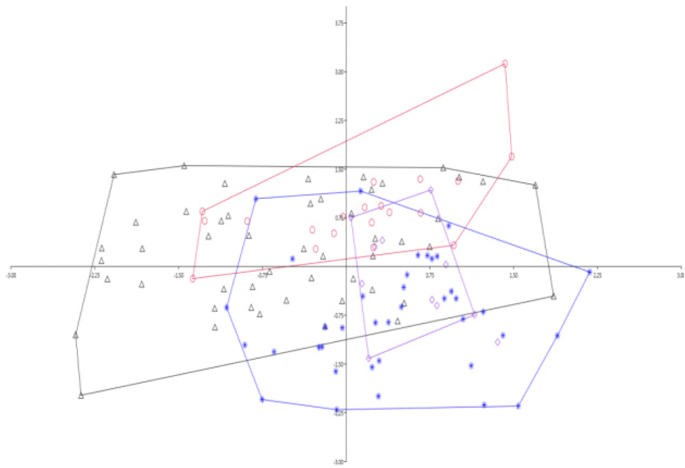
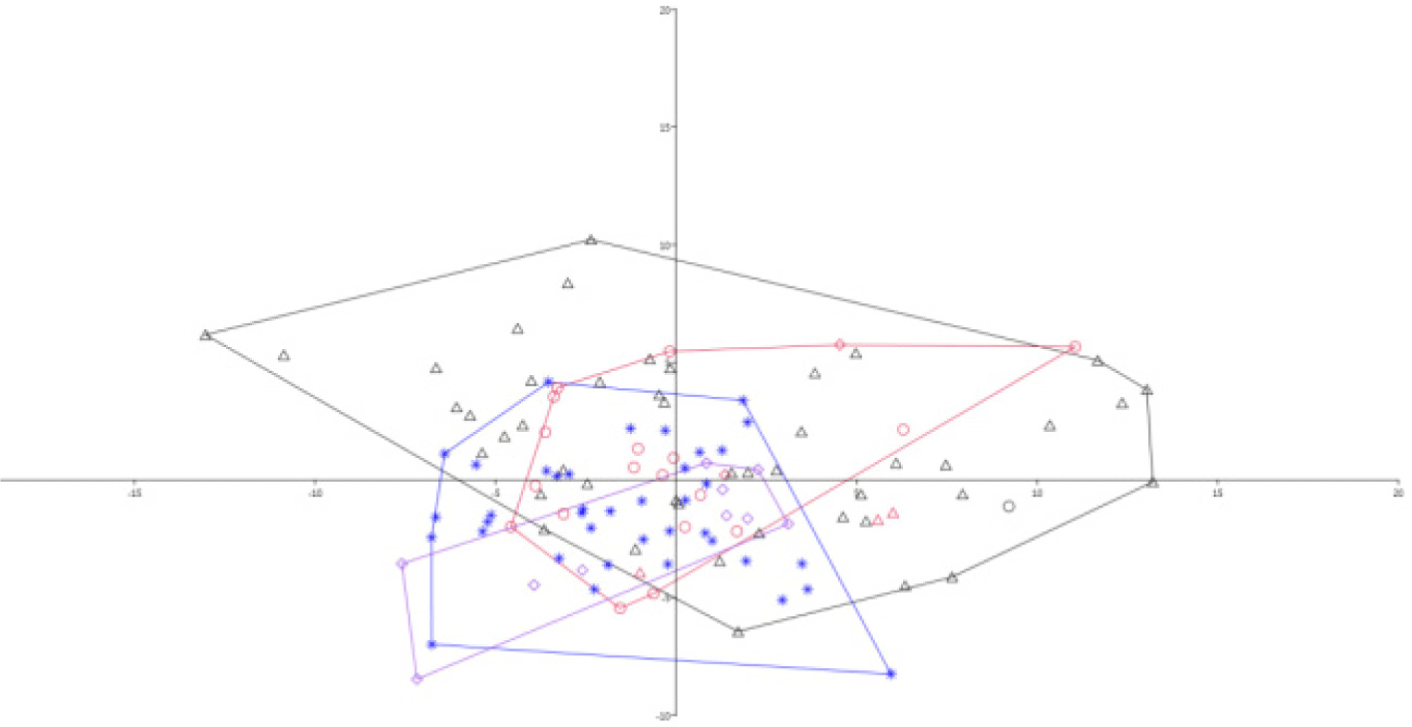
Accordingly, all the measurements used for this study were in comparison with percentages of SL and HL (Table 4). The PCA2 is mainly defined by pelvic fin length, followed by chin lobe length, head width, and snout length (Table 4 and Fig. 3). On the other hand, the third axis (PCA3) is mainly defined by head width, inner orbital width, post orbital length, and distance between pectoral fin length and anal fin length. The analysis was also made separately for each locality, and it showed the same distribution on the plane (Figs. 3 and 4). A comparison was also made between Nigeria and Congo, but no clear separation was apparently observed.

Table 4. Principal component analysis correlation for 29 morphological characters in percentages of specimens of Gnathonemus petersii (n = 119) according to the first four axes
Characters PCA1 PCA2 PCA3 PCA4
BD%SL 0.100327 –0.20547 –0.11291 –0.051338
HL%SL –0.055803 –0.13953 0.027644 0.03144492
PDD%SL –0.007756 –0.043839 0.16969 –0.007377
PAD%SL –0.03157 –0.15988 0.2440005 0.035016
PPID%SL –0.019866 –0.1244449 0.1196 0.061112
PPcD%SL –0.02894488 –0.070792 –0.002801 0.073372
DFL%SL 0.103313 0.13383 –0.22481 –0.102
DFH%SL –0.017017 –0.005896 0.032284 –0.102844
AFL%SL 0.046542 0.092232 –0.11727 –0.11362
PIFL%SL 0.0014866 1,12E–02* 0.0018565 –0.020714
PcFL%SL –0.011596 –0.018938 0.017774 0.0333
D-PIF-AF%SL 0.466422 0.0078618 0.13405 –0.0284655
D-PcF-AF%SL 0.055623 –0.062373 0.31539* –0.19036
D-PcF-PIF%SL 0.032116 –0.03349 0.19096 –0.12789
D-DF-AF%SL 0.064444 –0.092 –0.047715 –0.038536
CPL%SL –0.037044 –0.040335 0.051702 0.039224
CPH%SL –0.008426 –0.023506 0.047281 0.007211
CPD%SL 0.001108 –0.027248 0.048743 3–0.039608
HD%HL 0.88079 –0.32861* –0.10603 0.19284
HW%HL 0.28741 0.13957 0.42363* –0.65077
SnL%HL 0.093851 0.21127* 0.26092 0.035043
ED%HL –0.070074 0.038237 0.20123 0.19198
IoW%HL –0.008778 0.064079 0.40923* –0.65077
PoL%HL 0.094539 0.13839 0.39601* 0.61776
DNN%HL 0.030361 0.08125 0.033763 0.030726
DNE%HL 0.11376 0.087349 –0.028255 –0.043958
MW%HL 0.042283 0.10084 0.029503 0.055463
CIL%HL 0.24847 0.79109* -0.16825 0.076829

* The most important loadings.

PCA, principal component analysis; PCA1, first axis; PCA2, second axis; PCA3, third axis; PCA4, fourth axis; SL, standard length; BD, body depth; HL, head length; PDD, predorsal distance; PAD, preanl distance; PPID, prepelvic distance; PPcD, prepectoral distance; DFL, dorsal fin length; DFH, dorsal fin height; AFL, anal fin length; PIFL, pelvic fin length; PcFL, pectoral fin length; D-PIF-AF, distance between pelvic and anal fin; D-PcF-AF, distance between pectoral and anal fin; D-PcF-PIF, Distance between pectoral and pelvic fin; D-DF-AF, distance between dorsal and anal fin; CPL, caudal peduncle length; CPH, caudal peduncle height; CPD, caudal peduncle depth; HD, head depth; HW, head width; SnL, snout length; ED, eye diameter; IoW, inter orbital width; PoL, post orbital length; DNN, distance between nostrils; DNE, distance between nostril and eye; MW, mouth width; CIL, chin lobe length.
Download Excel Table
fas-28-6-372-g3
Fig. 3. A PCA (x axis- PCA1 and y axis- PCA2) of log transformed measurements (n = 119); (triangle- Congo, circle-Wouri, diamond- Sanaga, and star- Nigeria). PCA, principal component analysis; PCA1, first axis; PCA2, second axis.
Download Original Figure
fas-28-6-372-g4
Fig. 4. PCA in convexhaull of Gnathonemus petersii on percentage morphometrics (triangle-Congo, circle-Wouri, diamond-Sanaga, and star-Nigeria). PCA, principal component analysis.
Download Original Figure

In the present study, the test for clear separation analysis applied for the G. petersii species using morphometric measurements showed somehow separations in between Congo and Sanaga specimens (Fig. 4). The taxonomic description of a species has commonly relied on the description of unique sets of morphological characters. The morphometric measurements, which represent %SL and %HL, were the most discriminate characters used in the present analysis (Table 4; Figs. 3 and 4). Specific to the long-term morphology of fishes, morphological variation that is correlated with physiological parameters such as body size or sex can be directly assessed from measurement or dissection of preserved specimens and analyzed by collection year (Jacquemin & Pyron, 2016). In this particular aspect, natural history museums like the RMCA play a significant role in fish taxonomy studies not accessible for molecular and genetic studies (Biswal et al., 2018). Morphological variation studies are also important to adopt management implications in addition to solving taxonomical uncertainties (Stange et al., 2018). This is because morphological variations triggered by habitat differences are typically manifested in freshwater ecosystems, which are vulnerable to change in global climate impacts (Biswal et al., 2018).

Conclusion

This study assessed the morphological variation of G. petersii specimens collected from west and central African rivers. The morphology of fish has been the key source for taxonomic studies and has been able to clearly showed variations among fish. However, the current study could not clearly show differences among the different voucher specimens of G. petersii collected from different water bodies. Accordingly, ichthyologists recommend the use of genetic markers for verification before drawing conclusion on relationships among morphologically assessed varieties. The present finding is important to make revisions on the diverse African mormyrids.

Competing interests

No potential conflict of interest relevant to this article was reported.

Funding sources

Not applicable.

Acknowledgements

The author is indebted to appreciate all the 2017 trainees of FishBase and Taxonomy at RMCA for their support during data curation.

Availability of data and materials

Upon reasonable request, the datasets used in this study can be made available from the corresponding author.

Ethics approval and consent to participate

Informed consent was obtained from the RMCA/Ichthyology Unit to use the specimens for this study and ethically approved for research under Ethical Review Committee (Ref No. VM/RMCA/fb01/fb2017).

References

1.

Abdurahman SW, Ambak MA, Sheriff SM, Seah YG, Mohamed AA, Chowdhury AJK. Morphological variations of Plicofollis species (Siluriformes: Ariidae) in Peninsular Malaysia: an insight into truss network approach. Sains Malays. 2016; 45:1-7.

2.

Ahirwal SK, Singh J, Sarma K, Kumar T, Bharti V, Kumar A. Morphometric characteristics, length-weight relationships, and condition factors of five indigenous fish species from the river Ganga in Bihar, India. J Appl Ichthyol. 2023; 2023:1329222

3.

Alves-Gomes J, Hopkins CD. Molecular insights into the phylogeny of mormyriform fishes and the evolution of their electric organs. Brain Behav Evol. 1997; 49:343-51

4.

Bass AH. Electric organs revisited: evolution of a vertebrate communication and orientation organ.In: In: Bullock TH, Heiligenberg W, editors.editors Electroreception. New York, NY: John Wiley & Sons. 1986; pp p. 13-70.

5.

Bell CC. Sensory coding and corollary discharge effects in mormyrid electric fish. J Exp Biol. 1989; 146:229-53

6.

Biswal JR, Singh RK, Lal KK, Mohindra V, Kumar R, Kumar RG, et al. Molecular and morphological evidences resolve taxonomic ambiguity between Systomus sarana sarana (Hamilton, 1822) and S. sarana subnasutus (Valenciennes) and suggest elevating them into distinct species. Mitochondrial DNA B. 2018; 3:838-44

7.

Boden G, Teugels GG, Hopkins CD. A systematic revision of the large-scaled Marcusenius with description of a new species from Cameroon (Teleostei; Osteoglossomorpha; Mormyridae). J Nat Hist. 1997; 31:1645-82

8.

Brosse S, Charpin N, Su G, Toussaint A, Herrera-R GA, Tedesco PA, et al. FISHMORPH: a global database on morphological traits of freshwater fishes. Glob Ecol Biogeogr. 2021; 30:2330-6

9.

Carlson BA, Arnegard ME. Neural innovations and the diversification of African weakly electric fishes. Commun Integr Biol. 2011; 4:720-5

10.

Costa JL, de Almeida PR, Costa MJ. A morphometric and meristic investigation of Lusitanian toadfish Halobatrachus didactylus (Bloch and Schneider, 1801): evidence of population fragmentation on Portuguese coast. Sci Mar. 2003; 67:219-31

11.

Engelmann J, Nöbel S, Röver T, von der Emde G. The Schnauzenorgan-response of Gnathonemus petersii. Front Zool. 2009; 6:21

12.

Eschmeyer WN, Fricke R, van der Laan R. Catalog of fishes: genera, species, references [Internet]. 2020 [cited 2024 Sep 16]http://researcharchive.calacademy.org/research/ichthyology/catalog/fishcatmain.asp.

13.

Froese R, Pauly D. FishBase [Internet]. 2019 [cited 2023 Sep 18]http://www.fishbase.org.

14.

Gelsano ML, Demayo CG. Population variability in Poecilia reticulata based on morphological character measurements, length-weight and length-length relationships. AACL Bioflux. 2022; 15:1355-70.

15.

Günther A. Eine neue art von Mormyrus. Arch Naturgesch. 1862; 28:64

16.

Haryono H. Freshwater fishes of Pulosari mountain areas in Pandeglang Regency, Indonesia. BIO Web Conf. 2020; 19:00022

17.

Hubbs CL, Lagler KF. Fishes of the Great Lakes region. Ann Arbor, MI: University of Michigan Press. 1958

18.

Jacquemin SJ, Pyron M. A century of morphological variation in Cyprinidae fishes. BMC Ecol. 2016; 16:48

19.

Kerschbaumer M, Sturmbauer C. The utility of geometric morphometrics to elucidate pathways of cichlid fish evolution. Int J Evol Biol. 2011; 2011:290245

20.

King M. Fisheries biology, assessment and management. 2nd ed Oxford: Blackwell. 2007

21.

Lagler KF, Bardach JE, Miller RR. Lchthyology, the study of fishes. 2nd ed New York, NY: John Wiley & Sons. 1977.

22.

Lavoué S, Sullivan JP, Hopkins CD. Phylogenetic utility of the first two introns of the S7 ribosomal protein gene in African electric fishes (Mormyroidea: Teleostei) and congruence with other molecular markers. Biol J Linn Soc. 2003; 78:273-92

23.

Miller DJ, Lea RN. Guide to the coastal marine fishes of California. Calif Fish Bull. 1972; 157:1-235.

24.

Moyle PB, Cech JJ. Fishes: an introduction to ichthyology. 5th edEnglewood Cliffs, NJPrentice Hall 2000.

25.

Murta AG. Morphological variation of horse mackerel (Trachurus trachurus) in the Iberian and North African Atlantic: implications for stock identification. ICES J Mar Sci. 2000; 57:1240-8

26.

Nelson JS, Grande TC, Wilson MVH. Fishes of the world. 5th edHoboken, NJJohn Wiley & Sons 2016

27.

Nwani CD, Odoh GE, Ude EF, Okogwu OI. Food and feeding habits of Gnathonemus petersii (Osteichthyes: Mormyridae) in Anambra river, Nigeria. Int Aquat Res. 2011; 3:45-51.

28.

Simanovsky S, Medvedev D, Tefera F, Golubtsov A. First cytogenetic information for five Nilotic elephant fishes and a problem of ancestral karyotype of the family Mormyridae (Osteoglossiformes). Comp Cytogenet. 2020; 14:387-97

29.

Stange M, Aguirre-Fernández G, Salzburger W, Sánchez-Villagra MR. Study of morphological variation of northern Neotropical Ariidae reveals conservatism despite macrohabitat transitions. BMC Evol Biol. 2018; 18:38

30.

Strauss RE, Bond CE. Taxonomic methods: morphology.In: In: Moyle P, Schreck C, editors.editors Methods for fish biology. Bethesda, MD: American Fisheries Society. 1990; pp p. 109-140.

31.

Sullivan JP, Lavoué S, Hopkins CD. Discovery and phylogenetic analysis of a riverine species flock of African electric fishes (Mormyridae: Teleostei). Evolution. 2002; 56:597-616