Introduction
The sex determination of teleost fishes falls into two functional categories: gonochorism and hermaphroditism. In gonochoristic species, the sex established during sexual differentiation remains fixed throughout the lifetime of the organism. In contrast, hermaphroditic species exhibit sexual plasticity, allowing for sex change from the initially determined sex to the opposite sex. Hermaphroditism is further classified into protandrous hermaphroditism, where the transition occurs from male to female, and protogynous hermaphroditism, where the transition occurs from female to male. In teleost fishes, environmental and social factors primarily govern sex change. Research on protogynous hermaphroditic fish in the family Labridae has shown that when the dominant male is removed from the group, the largest remaining female undergoes sex change to become male (Thomas et al., 2019; Todd et al., 2019). Conversely, in clownfish, which exhibit protandrous hermaphroditism, the removal of the dominant female triggers sex change in the largest subordinate male, which then becomes the new dominant female (Casas et al., 2016; Parker et al., 2024). Once sex change is initiated in fish, the reproductive endocrine system transitions to express characteristics of the opposite sex.
The reproductive endocrinology of fish is regulated by the hypothalamus-pituitary-gonad (HPG) axis, where gonadotropin-releasing hormone (GnRH), gonadotropins (GtHs), and sex steroid hormones are secreted by each organ to coordinate reproductive endocrine functions. While testosterone is generally recognized as an androgenic hormone characterizing male traits in mammals (Preston et al., 2012), it exhibits bipotentiality in teleost fish, influencing both males and females (Goymann & Wingfield, 2014). The sex steroid hormones 11-ketotestosterone (11-KT) and estradiol-17β mediate androgenic and estrogenic functions (Jang & Ji, 2015). Testosterone secreted by Leydig cells in the gonads is ultimately converted into the androgenic hormone 11-KT by 11β-hydroxylase (Golshan & Alavi, 2019). When testosterone is metabolized by aromatase, it is converted into estradiol (E2), a C18 compound with estrogenic activity. In this process, the C19 methyl group (–CH₃) is removed, and the A-ring undergoes aromatization, resulting in an aromatic molecular structure. Through this oxidation reaction, the ketone group of testosterone is converted into a hydroxyl group (–OH), resulting in the formation of E2 (Diotel et al., 2010). Consequently, increased 11-KT secretion promotes testicular development and masculinization (Thomas et al., 2019), while elevated E2 secretion leads to ovarian development and feminization (Casas et al., 2016). Among teleosts, hermaphroditic species include members of the families Labridae, Serranidae, Pomacanthidae, Sparidae, as well as hamlet fishes, anemonefishes, and ribbon eels. Within this diverse group, the Serranidae family exhibits protogynous hermaphroditism.
Serranidae are emerging as economically important aquaculture species, particularly in tropical and subtropical regions such as Southeast Asia (Kawabe & Kohno, 2009). As of 2017, global aquaculture production of groupers reached approximately 183,989 metric tons, representing a 230% increase compared to 2010 (FAO, 2017). According to FAO (2020), production further increased to around 200,000 tons in 2018. Groupers have garnered attention as promising aquaculture species capable of withstanding elevated water temperatures associated with global climate change. Notably, the hybrid Epinephelusmoara ♀ × Epinephelus lanceolatus ♂ has gained significant interest from the aquaculture industry due to its rapid growth and resistance to high water temperatures. These hybrids exhibit accelerated growth under high-temperature conditions, enabling size control for market demands and year-round production within land-based aquaculture systems (Kim et al., 2020). However, the time required for sex change is considerably longer for the fish in the Serranidae family. The onset of first puberty and functional female maturation is typically followed by sex change, which generally occurs over a period of 3 to 7 years (Bouchereau et al., 1999; Park, 2021). Consequently, sourcing wild male broodstock for breeding purposes remains difficult and poses considerable risks. Recent research efforts have therefore focused on inducing precocious sex change to address this limitation (Wang et al., 2017). The extended time frame for natural sex change significantly impacts aquaculture profitability. Producers must maintain breeding stocks for several years before functional males become available, which increases operational costs and reduces breeding efficiency. For artificial sex change, 17α-methyltestosterone (MT) is commonly used. MT is a synthetic androgen that induces artificial masculinization by promoting female-to-male sex change in teleost fish (Han et al., 2024; Hu et al., 2011; Liu et al., 2023; Wang et al., 2017). As an exogenous androgenic compound, MT functions similarly to the endogenous 11-KT hormone and exerts regulatory effects on the HPG axis (Liu et al., 2023). Artificial administration of MT suppresses the secretion of GnRH in the hypothalamus, as well as follicle-stimulating hormone (FSH) and luteinizing hormone (LH) in the pituitary gland, thereby reducing the synthesis of sex steroid hormones in both testes and ovaries (Liu et al., 2023). In particular fish species, MT treatment has been shown to inhibit aromatase (cyp19a1) activity, consequently reducing the conversion of testosterone into E2 and suppressing ovarian development in female individuals (Garcia et al., 2013). However, MT-induced masculinization has been associated with adverse side effects, including impaired reproductive function, growth retardation, and endocrine disruption (Boudreau et al., 2005; Tan-Fermin et al., 1994). Therefore, to achieve stable and safe sex change in aquaculture species, further research is required to identify alternative sex changing agents that can effectively replace exogenous hormones such as MT.
Recently, studies have been conducted to induce sex change using endogenous hormones such as aromatase inhibitors (AI) as alternatives to MT (Bhandari et al., 2005; Goikoetxea et al., 2021; Nozu et al., 2009). AI-induced masculinization has been observed in groupers, including the dusky grouper (Epinephelus marginatus), greasy grouper (Epinephelus tauvina), leopard coral grouper (Plectropomus leopardus), and the orange-spotted grouper (Epinephelus coioides) (Garcia et al., 2013; Huang et al., 2019; Miyoshi et al., 2025; Ranjan et al., 2015). In teleost fish, masculinization has also been reported following administration of letrozole (a compound belonging to class AI) in fish species such as Nile tilapia (Oreochromis niloticus), rainbow trout (Oncorhynchus mykiss), and largemouth bass (Micropterus salmoides) (Alijani et al., 2022; Betancur López et al., 2014; Zhang et al., 2024). Unlike direct-acting androgens, AI compounds exert their effects indirectly by disrupting estrogen synthesis rather than supplying exogenous hormones. Letrozole functions as a potent AI, blocking the enzymatic conversion of androgens into estrogens, a key biochemical step required for maintaining ovarian development. Suppression of aromatase activity leads to a marked reduction in endogenous estrogen levels, thereby inhibiting ovarian growth and function. As estrogen signaling declines, androgen dominance is enhanced, shifting the endocrine balance toward masculinization (Irani & Noori, 2023). This hormonal reprogramming suppresses female differentiation pathways and promotes the activation of testicular development and male-specific gene expression, ultimately steering gonadal differentiation toward a male phenotype (Zhang et al., 2024). In Korea, few reports exist on artificial sex change in longtooth grouper (Epinephelus bruneus), and the underlying mechanisms of sex change remain poorly understood. Considering the economic importance of longtooth grouper in South Korean aquaculture, the development of a reliable technique for stable female-to-male sex change is essential for the successful breeding. To date, no study has been conducted to compare the effect of administration of letrozole and MT on sex change and the expression of GtHs and sex steroid hormone receptor genes in longtooth grouper. Therefore, in this study, both an endogenous hormone (AI) and an exogenous hormone (MT) were administered to induce sex change in longtooth grouper, and the masculinization process was examined through histological analysis of the gonads. Furthermore, to investigate reproductive endocrine changes in the pituitary and gonads during the hormone-induced masculinization process, mRNA expression levels of pituitary GtHs and gonadal sex steroid hormone receptors were analyzed.
Materials and Methods
The female longtooth grouper used in the experiment were reared at the Jeju Fisheries Research Institute, National Institute of Fisheries Science, Korea. A total of 78 similar-sized juvenile fish (body weight, 404 ± 43.1 g; total length, 29.4 ± 0.9 cm) were randomly distributed into three separate fiber-reinforced plastic tanks (5,000 L) with 26 fish in each. All fish were maintained in a common flow-through system with uniform filtered seawater flow (20 L/min) and water quality conditions for 12 weeks. Water temperature (23.3°C ± 1.5°C), dissolved oxygen (6.34 ± 0.61 mg/L), pH (8.12 ± 0.2), and salinity (31.3 ± 1.2 ppt) were monitored and maintained within the same range across all tanks. A controlled photoperiod of 12 h light:12 h dark was applied using artificial lighting (202 lx). Tanks were positioned to minimize spatial heterogeneity and reduce the influence of extraneous environmental factors. Commercial feed (crude protein, 52%; crude lipid, 11%; Suhyup Feed, Uiryeong, Korea) was provided twice daily during the experimental period. MT (17α-methyltestosterone) was purchased from Sigma–Aldrich (St. Louis, MO, USA), and the AI (letrozole) was purchased from Femara, Novartis (Basel, Switzerland). Fish in two experimental groups (MT and AI) were administered intramuscular injections of MT and AI at 5 mg/kg body weight following the method used in Hur et al. (2012). Before administration, hormones were dissolved in an ethanol and coconut oil mixture (1:5 v/v), which was used as the hormone carrier. The control group received intramuscular injections of the ethanol and coconut oil mixture (1:5, v/v) at a dosage of 5 mg/kg body weight, matching the administration protocol of the hormone-treated groups. Hormone administration was carried out at 0, 3, and 6 weeks of the experiment, and samples were collected at 3, 6, and 12 weeks. From each treatment group, five fish were randomly selected at each time point for sample collection, and they were anaesthetized with 0.05% (500 ppm) tricaine methanesulfonate (MS-222, Sigma-Aldrich) before sampling. Those five fish were sacrificed by decapitation, and the pituitary and gonads were removed. Sampling was destructive, and different individuals were used at each sampling time point. The collected gonad samples were weighed for calculation of the gonadosomatic index (GSI = gonad weight × 100 / body weight) and fixed in Bouin’s solution for histological observation of changes in gonadal characteristics. Histology slides were prepared, stained, and observed according to the methods described in Medagoda et al. (2025). To investigate the expression changes in the GtHs and sex steroid hormone receptors, the whole brain, pituitary, and parts of the gonad tissue were collected, immediately frozen in liquid nitrogen, and stored at –80°C until real-time quantitative polymer chain reaction (RT-qPCR) analysis. The expression levels of key neuroendocrine genes, including kisspeptin 1 (Kiss1), kisspeptin receptor (GPR54), and gonadotropin-releasing hormone (GnRH), gonadotropin genes, including follicle-stimulating hormone beta subunit (fshβ) and luteinizing hormone beta subunit (lhβ), and sex steroid hormone receptors, including androgen receptor (ar), estrogen receptor alpha (erα), estrogen receptor beta 1 (erβ1), and estrogen receptor beta 2 (erβ2), were evaluated. For cloning and determining tissue-specific distribution of the sex steroid hormone receptor in the longtooth grouper, five fish were sampled after being anaesthetized with 0.05% MS-222. Whole brain, pituitary, and gonad tissues were extracted. The extracted samples were immediately frozen in liquid nitrogen and stored at –80°C.
For gene cloning of sex steroid hormone receptors, a degenerated primer set was designed based on regions of high identity from the nucleotide sequences of these receptors in other fish. cDNAs of the sex steroid hormone receptors of longtooth grouper were amplified by RT-PCR using a degenerate primer set (Table 1). PCR products were purified using a DNA purification kit (Promega, Madison, WI, USA), and purified DNA fragments were cloned into the pGEM-T Easy Vector System (Promega). Sequencing was conducted using PRISM 3730XL Analyzer (Applied Biosystems, Waltham, MA, USA). The obtained nucleotide and amino acid sequences were analyzed using the BLASTN program (National Center for Biotechnology Information, National Institutes of Health, Bethesda, MD, USA) and the ORF finder program (National Center for Biotechnology Information, National Institutes of Health; https://www.ncbi.nlm.nih.gov/orffinder). Multiple alignment and homology of obtained amino acids were analyzed using the Clustal Omega software (https://www.ebi.ac.uk/jdispatcher/msa/clustalo), and information on other fish species used in the analysis is shown in Table 2. A phylogenetic tree was constructed using 1,000 bootstrap trials by the Neighbor-Joining method using pairwise sequence comparisons (pairwise deletion of gaps) in Molecular Evolutionary Genetics Analysis (MEGA) v.11 software.
RT-PCR, real-time polymer chain reaction; RT-qPCR, real-time quantitative polymer chain reaction; ar, androgen receptor; erα, estrogen receptor alpha; erβ1, estrogen receptor beta 1; erβ2, estrogen receptor beta 2; fshβ, follicle-stimulating hormone beta subunit; lhβ, luteinizing hormone beta subunit; Kiss1, kisspeptin1; GPR54, kisspeptin receptor; and GnRH, gonadotropin-releasing hormone.
The brain and pituitary tissues were thoroughly homogenized with TriPure Isolation reagent (Roche Diagnostics, Indianapolis, IN, USA), and total RNA was extracted following the manufacturer’s protocol. The extracted total RNA was treated using the RQ1 RNase-Free DNase (Promega) to prevent genomic DNA contamination. cDNA was synthesized using the Prime ScriptTM RT Reagent kit (TaKaRa Bio, Kusatsu, Japan) and was used for gene cloning analysis, RT-PCR, and RT-qPCR.
Gene-specific primer sets of 500–600 bp were designed based on the sequences of each gene obtained by cloning (Table 1). PCR amplification was performed using EmeraldAmp PCR Master Mix (12.5 μL master mixture per 25 μL reaction) (TaKaRa Bio), 10 µL of primers, and 50 ng of cDNA for 28 cycles of denaturation (45 s, 94°C), annealing (45 s, 58°C), and extension (1 min, 72°C). PCR products were identified using electrophoresis of ethidium bromide on 2% agarose gel and visualized on a UV transilluminator (WSE-5200 Printgraph 2M, Bio-Rad, Hercules, CA, USA).
Gene expression was analyzed using RT-qPCR. The analysis was performed using the CFX96TM Real-time System (BioRad) and LightCycler 480 SYBR Green I Master (Roche Diagnostics GmbH, Mannheim, Germany). Primer sets were designed to be approximately 80–150 bp using the Primer-BLAST program (http://www.ncbi.nlm.nih.gov/tools/primer-blast/) as shown in Table 1. Primer efficiency was evaluated using a standard curve generated from a serial dilution of cDNA. The amplification efficiency for all primer pairs ranged between 90% and 110%, with correlation coefficients (R²) greater than 0.99, indicating acceptable amplification performance for RT-qPCR analysis. RT-qPCR reaction mixture contained 25 µl of SYBR Green Mix, 10 µL of primers, and 20 ng of cDNA. The PCR was carried out for 40 cycles, each comprising denaturation at 94°C for 45 s, annealing at 58°C for 45 s, and extension at 72°C for 1 min. Gene expression was quantified by absolute RT-qPCR using standard curves generated from serial dilutions of standard DNA, and the expression was normalized to the reference gene β-actin. β-actin was selected as the internal reference gene because of its stable and consistent expression in teleost fish, including grouper, gonadal, and pituitary tissues under hormonal manipulation.
All data were presented as box-and-whisker plots, and all statistical analyses were performed using GraphPad Prism software (version 8.0.1, San Diego, CA, USA). All data sets were tested for normality using the Shapiro–Wilk test. The experimental data were subjected to two-way analysis of variance (ANOVA) followed by Tukey’s multiple comparison test. Statistical significance was accepted when p < 0.05.
Results
Partial cDNA sequences encoding sex steroid hormone receptors (ar, erα, erβ1, and erβ2) were successfully amplified from longtooth grouper pituitary tissue. The obtained sequences were 987 bp (329 aa) for ar, 784 bp (262 aa) for erα, 697 bp (232 aa) for erβ1, and 987 bp (328 aa) for erβ2. Sequence analysis revealed 100% amino acid identity between longtooth grouper and orange-spotted grouper for all four receptors (Fig. 1A and 1B). Furthermore, comparative analysis with other teleost species showed high sequence conservation with homology ranging from 92.4%–93.0% for ar, 88.9%–91.6% for erα, 81.5%–86.6% for erβ1, and 91.2%–92.1% for erβ2.
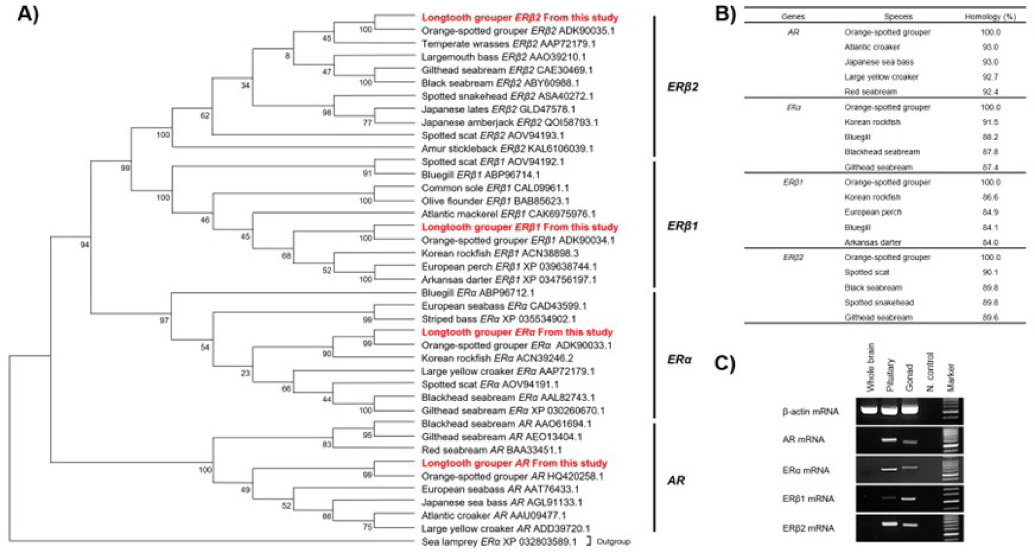
The tissue distribution of sex steroid hormone receptors (ar, erα, erβ1, and erβ2) was examined by RT-PCR across three tissues, including whole brain, pituitary, and gonad, and expressions were visualized in Fig. 1C. The ar was strongly expressed in the pituitary and weakly in the gonad. The erα was expressed in all tissues except the whole brain, and the strongest expression was observed in the pituitary. The erβ1 was detected in all tissues except the whole brain, with particularly strong expression in the gonad. The erβ2 showed the most restricted expression pattern, being absent from the whole brain but strongly expressed in the pituitary and gonad. All four receptors showed consistent expression in the pituitary and gonad, while no expression was detected in the brain.
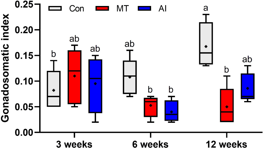
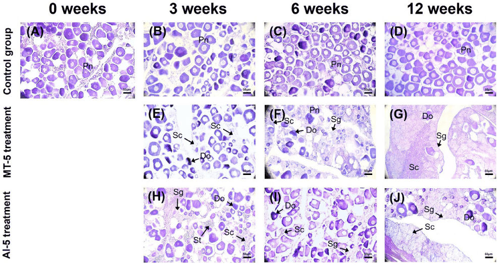
Both MT and AI treatments effectively induced masculinization in female longtooth grouper, as demonstrated by changes in GSI and histomorphological observations of gonads over the 12-week experimental period (Figs. 2 and 3). The control group showed a significant increase in GSI compared to the initial time point (p < 0.05) (Fig. 2). In contrast, both MT and AI treatments suppressed gonadal development. The MT group showed significantly lower GSI than the control at week 6 and 12 (p < 0.05), whereas the AI group exhibited significantly reduced GSI only at week 6 (p < 0.05). Notably, GSI levels in both treatment groups remained comparable to initial values throughout the experiment (p > 0.05). Two-way ANOVA revealed that there was a significant interaction effect between hormonal administration and time (p = 0.0063). Histological examination revealed that fish in the control group maintained their ovarian structure containing perinucleolar oocytes (Pn) throughout the 12-week experimental period, showing no evidence of sex change (Fig. 3A–3D). In the MT group, masculinization became evident (partial sex change) from the third week with the appearance of degenerating oocytes (Do) and spermatocytes (Sc) alongside Pn, followed by the emergence of spermatogenic cells, including spermatogonia (Sg) and Sc at 6 weeks (Fig. 3E and 3F). By 12 weeks, MT-treated gonads contained extensive populations of Sg and Sc with numerous Do (Fig. 3G). The AI treatment induced similar but more rapid masculinization, with a higher number of Do and early spermatogenesis from the third week (Fig. 3H and 3I). At the end of the 12th week of the experiment, the gonads of AI-treated fish showed fewer Pn and Do, along with substantial numbers of Sg and Sc (Fig. 3J).
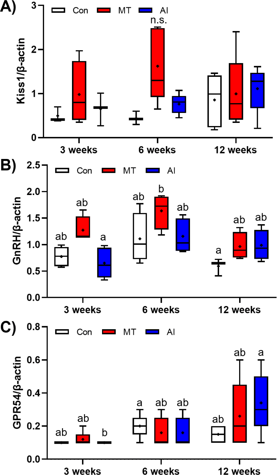
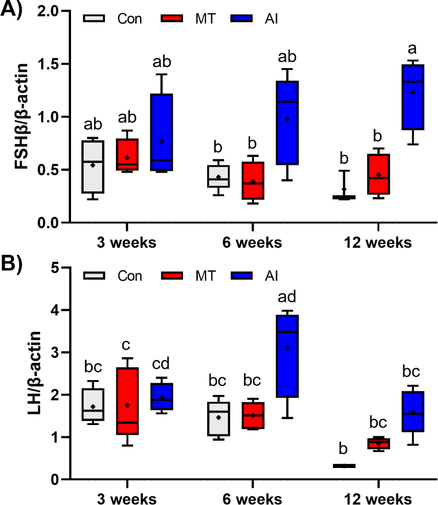
Hormone-induced changes in the expression of pituitary GtHs and gonadal sex steroid hormone receptors are illustrated in Figs. 4–6. In the whole brain, Kiss1 and gnrh did not differ significantly in the control, MT, and AI groups (p > 0.05) (Fig. 4A and 4B). However, gpr54 expression was significantly upregulated in the AI group at 12 weeks compared to the expression at 3 weeks (p < 0.05) (Fig. 4C). No significant interaction effect between hormonal treatments and time was observed for the expression of Kiss1(p= 0.3871), gnrh(p= 0.1582) and gpr54 (p = 0.2105). Pituitary fshβ and lhβ expression remained stable in both control and MT groups throughout the 12-week experimental period (Fig. 5A and5B). In contrast, AI treatment significantly upregulated fshβ gene expression at 12 weeks and lhβ gene expression at 6 weeks compared with the 12-week control group (p < 0.05). Two-way ANOVA showed no significant interaction between hormone injection and time on the gene expressions of fshβ (p = 0.1220) and lhβ (p = 0.0802).
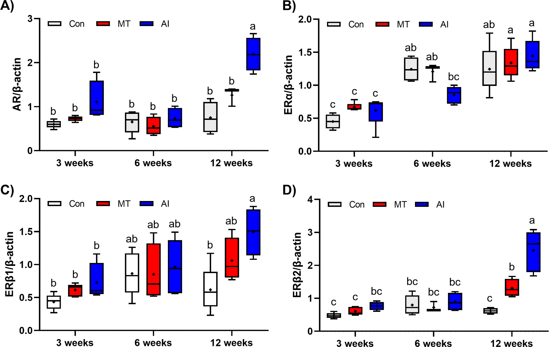
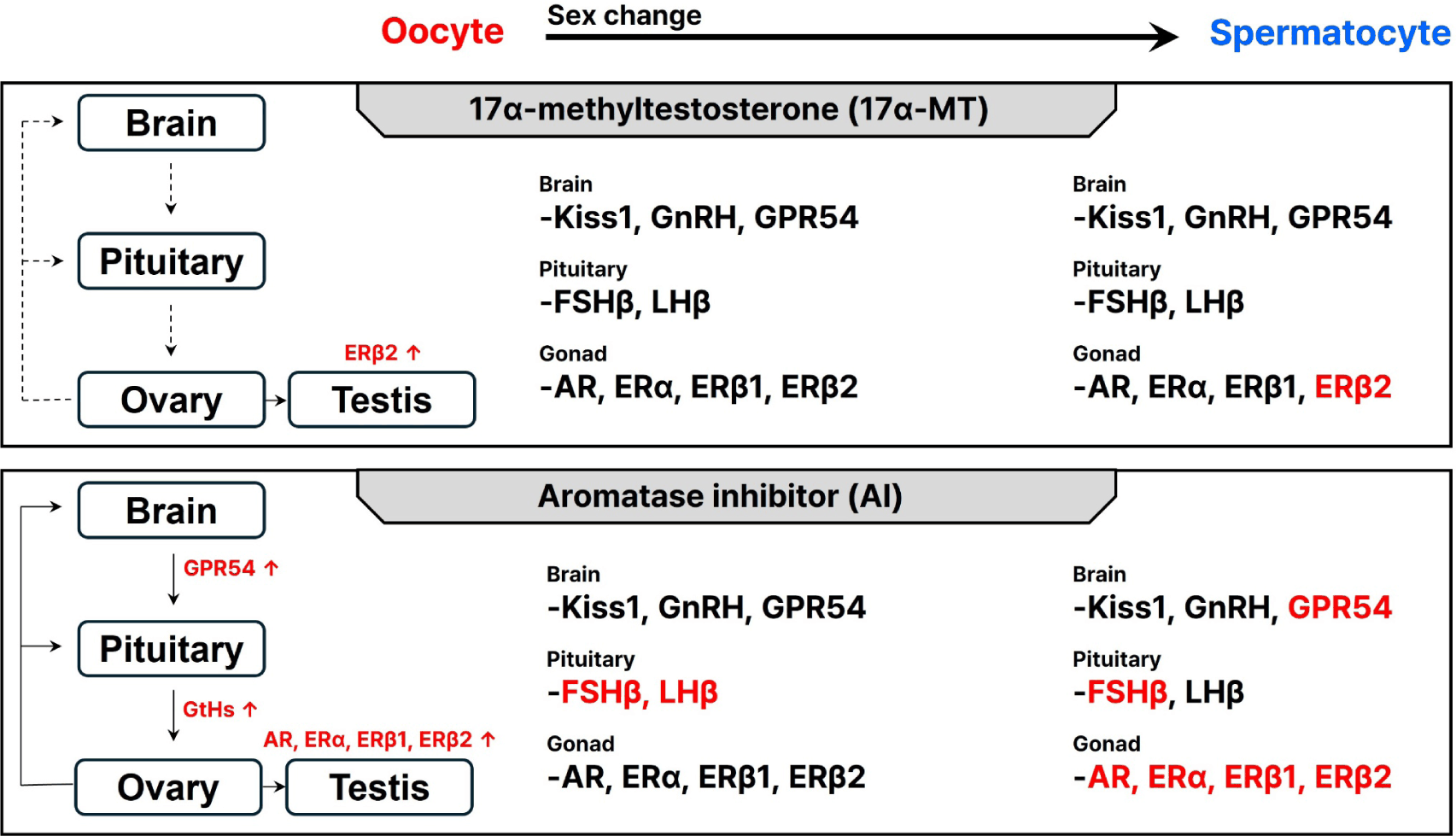
MT treatment had minimal effects on sex steroid hormone receptor expression in gonadal tissue, and the expression of ar, erα, and erβ1 showed no significant differences compared to control treatment throughout the experimental period (p > 0.05) (Fig. 6A–6D). The expression of the ar gene was significantly enhanced in the AI group compared to the control treatment at week 12, demonstrating its pronounced effect on the expression of gonadal sex steroid hormone receptors (p < 0.05) (Fig. 6A). A significant interaction between hormonal treatment and time was observed for ar gene expression (p = 0.0036). The expression of the erα gene in the AI group was significantly decreased at 6 weeks compared to the control treatment (p < 0.05). The erα showed no significant difference compared to the control treatment at 12 weeks (Fig. 6B). Two-way ANOVA showed a significant interaction between hormone injection and time on the gene expressions of erα (p = 0.0245). erβ1 expression was significantly elevated in the MT group compared to controls at week 12 (p< 0.05) (Fig. 6C). Moreover, erβ2 gene expression was significantly higher in the AI group compared to both the control and MT groups at week 12 (p < 0.05) (Fig. 6D). No significant interaction between hormonal treatment and time was observed for the expression of erβ1 (p = 0.1185), whereas the gene expression of erβ2 showed a significant interaction effect (p = 0.0004). The modulatory effects of exogenous MT and AI on the brain-pituitary-gonadal (BPG) axis in longtooth grouper and possible underlying mechanisms are illustrated in Fig. 7.
Discussion
Our study investigated the early sex change and masculinization process in the hermaphroditic longtooth grouper through intrinsic and extrinsic hormone treatments. We analyzed changes in the gonadal developmental phase and the gene expression of GtHs and steroid hormone receptors. Partial sequencing of steroid hormone receptors in longtooth grouper revealed four receptor subtypes: ar, erα, erβ1, and erβ2. Partial nucleotide sequences of all receptors exhibited 100% homology with those of the orange-spotted grouper and relatively high homology with other fish species. Tissue-specific distribution analysis of the steroid hormone receptors showed that all receptors were expressed in various peripheral tissues except for the whole brain. Common transcriptional expression was observed in the pituitary and gonads. In fish, each steroid hormone receptor has been detected in the liver, where they are reported to participate in vitellogenesis (deVlaming et al., 1984; Johnson et al., 1991). In mature male sticklebacks (Gasterosteus aculeatus), the kidney hypertrophies are observed during nest-building for reproduction, coinciding with increased ar mRNA expression (Hoffmann et al., 2012). This suggests that ar may play a role in reproductive behaviors, likely by regulating protein synthesis in the kidney during the onset of breeding. Similarly, in rainbow trout, erαmRNA expression has been detected in various lymphoid organs, and its localization was confirmed in the skin and intestine through immunohistochemistry (Massart et al., 2014). These findings imply that era may also be involved in immune mechanisms. Based on these observations, it is hypothesized that the ar and ers identified in this study may also contribute to diverse physiological processes, including reproduction and sexual maturation. Our results demonstrated that both MT and AI treatments successfully induced masculinization in female longtooth grouper, as evidenced by the suppression of GSI increases and the appearance of male gonadal characteristics. The control group exhibited a significant GSI increase at 12 weeks, reflecting normal ovarian development, while both treatment groups maintained significantly lower GSI levels with no observable increase. Notably, histological analysis revealed the emergence of spermatogonia in treated fish ovaries as early as 3 weeks, progressing to testis-dominant gonads containing spermatogonia and spermatocytes by 12 weeks. These morphological changes confirm that both hormone treatments effectively disrupted normal female development and initiated male differentiation. The degree of masculinization varied between treatments and time points, with both MT and AI treatments promoting only partial, rather than complete, sex change by week 12. While spermatogenic cells were clearly present, residual oocytes remained in treated fish, indicating an intermediate transitional state rather than complete differentiation.
The GSI was increased in control fish, confirming that the experimental conditions supported typical ovarian development. This validates that the suppressed gonadal growth observed in the treated groups resulted specifically from hormone intervention rather than environmental stress or suboptimal rearing conditions. The mechanisms underlying these observed masculinization effects can be explained by the distinct but complementary actions of MT and AI on the reproductive endocrine system. Our MT treatment results align with established findings that MT functions through multiple pathways within the HPG axis, exerting both positive and negative feedback mechanisms (Han et al., 2024; Liu et al., 2023). The rapid appearance of male gonadal tissue observed in our study supports previous research demonstrating that MT induces apoptosis in ovarian tissues (Lee et al., 2017) while simultaneously upregulating testis-determining genes such as SRY-box transcription factor 9 (sox9) and doublesex and mab-3 related transcription factor 1 (dmrt1) (Bhat et al., 2021). Further, they explained that MT-induced upregulation of dmrt1 and sox9 activates the testis-specific transcriptional network that promotes Sertoli cell differentiation and spermatogenesis while suppressing ovarian gene expression and inducing follicular apoptosis, thereby driving rapid male gonadal differentiation. In contrast, AI treatment achieved similar masculinization outcomes through a different mechanism, supporting the hypothesis that blocking aromatase activity effectively shifts the hormonal balance toward androgenization. By preventing testosterone conversion to E2, AI treatment likely elevated endogenous androgen levels, which subsequently stimulated GnRH and gonadotropin secretion. This interpretation is consistent with previous studies in Barilius species, where increased LH levels promoted gonadal androgen synthesis and facilitated testicular development (Garcia et al., 2013). Notably, our gene expression analysis revealed that AI treatment uniquely affected pituitary fshβ and lhβ expression, while both treatments modulated gonadal steroid receptor expression, suggesting that AI may have broader effects on the central regulatory components of the HPG axis compared to MT. The differential effects of MT and AI treatments observed in our study are further supported by comparative research in related grouper species, which reveals the complex and species-specific nature of gonadotropin regulation during sex change. In orange spotted grouper, MT administration resulted in sustained upregulation of lhβ expression accompanied by elevated plasma 11-KT levels throughout the entire experimental period (Huang et al., 2019). Thus, letrozole induces masculinization by blocking the conversion of testosterone to E2, reducing E2-mediated negative feedback on HPG axis, increasing fshβ and lhβ expression, enhancing endogenous androgen production and androgen receptor signaling in the gonads, which suppresses ovarian development, reduces GSI, and promotes testicular differentiation.
These results suggest that MT treatment led to an increase in LH, which subsequently induced the biosynthesis of testosterone and 11-KT, both androgens, thereby triggering sex change (Hu et al., 2011). Similarly, research on the honeycomb grouper (Epinephelus merra) demonstrated that FSH, rather than LH, serves as the primary trigger for sex change. In here, fshβ expression remains elevated throughout the masculinization process, and FSH treatment successfully induces male-directed sex change alongside increased plasma 11-KT levels (Kobayashi et al., 2010). Interestingly, our findings in longtooth grouper showed that AI treatment uniquely affected both pituitary fshβ and lhβ expression, while MT treatment primarily influenced gonadal steroid receptor expression without affecting pituitary GtHs. This suggests that AI may exert broader regulatory effects on the central components of the HPG axis compared to MT. It further indicates the potential species-specific differences in gonadotropin sensitivity and sex change mechanisms among grouper species. These species-specific differences in gonadotropin regulation can be further understood within the broader context of hermaphroditic reproductive strategies. Comparative studies reveal distinct patterns between protogynous and protandrous hermaphrodites in their gonadotropin expression during sex change. In protandrous species such as black porgy (Acanthopagrus schlegelii) and cinnamon clownfish (Amphiprion melanopus), LH expression plays a pivotal role in initiating male-to-female sex change, with both fshβ and lhβ expression increasing during the transition, reaching highest levels in mature females (An et al., 2008; An et al., 2010). In contrast, protogynous species like the groupers show different regulatory patterns, where FSH expression markedly increases from the early phase of female-to-male sex change and remains elevated throughout the process, while LH shows minimal changes. Our findings in longtooth grouper, a protogynous hermaphrodite, align with this pattern, as AI treatment specifically upregulated both fshβ and lhβ expression, suggesting that artificial hormone manipulation can activate both gonadotropin pathways to achieve effective masculinization. This understanding of species-specific and strategy-specific gonadotropin regulation provides important insights for developing targeted sex change protocols in hermaphroditic fish species for aquaculture applications.
The differential gene expression patterns observed in our study provide insights into the distinct mechanisms underlying MT and AI-induced masculinization. Our findings revealed that MT treatment primarily affected gonadal steroid receptors, specifically upregulating erβ2 expression at 12 weeks without altering pituitary gonadotropin levels. In contrast, AI treatment induced broader endocrine changes, significantly increasing both pituitary fshβ and lhβ expression alongside gonadal ar, erβ1, and erβ2 expression. These results suggest that in longtooth grouper, gonadal steroid receptors may play a more direct role in mediating sex change than pituitary GtHs, with AI treatment affecting both central and peripheral components of the reproductive axis. The observed increase in AR expression following AI treatment aligns with previous studies demonstrating the importance of androgen signaling in sex change. In rainbow trout, AR regulates male reproductive function and can induce partial masculinization in ovarian tissue, although complete sex change may not always occur (Baron et al., 2007). The pronounced AR upregulation in our AI-treated fish may explain the effective masculinization observed histologically. Similarly, the differential expression of estrogen receptor subtypes supports their distinct roles in reproductive processes. In mandarin fish (Siniperca chuatsi), MT-induced sex change was associated with upregulation of multiple estrogen receptors and arβ, suggesting their involvement in spermatogenesis and masculinization, while erβ2 and erβ1 were linked to vitellogenesis and ovarian development (Han et al., 2024). The mechanisms underlying these treatments reflect their different approaches to hormonal manipulation. MT functions as a direct androgen agonist, binding to androgen receptors to activate male-specific gene pathways while simultaneously suppressing estrogen synthesis to prevent feminization (Wang et al., 2017). This direct androgenic action explains the observed masculinization despite minimal changes in gonadotropin expression. Conversely, AI treatment blocks aromatase (cyp19a1) activity, preventing testosterone conversion to E2 and creating a relative androgen excess that promotes testicular differentiation (Doering et al., 2021; Jiang et al., 2022). This mechanism maintains endogenous hormone production pathways, which may explain the broader effects on both pituitary and gonadal gene expression observed in our study. From an aquaculture perspective, both treatments successfully induced sex change in longtooth grouper, as evidenced by suppressed GSI values and the appearance of spermatogenic cells. However, the different endocrine profiles suggest that AI treatment may offer advantages over MT. AI-induced masculinization relies on endogenous hormone modulation rather than exogenous androgen supplementation, potentially resulting in a more physiological sex change process (Babiak et al., 2012; Gennotte et al., 2015). Given the documented side effects associated with MT treatment in aquaculture applications, including growth retardation and endocrine disruption, AI treatment may represent a safer and potentially more sustainable approach for producing male broodstock in longtooth grouper farming. An important consideration for aquaculture applications is whether AI-induced masculinization represents a permanent or reversible change. While our 12-week study period demonstrated sustained masculinization, the long-term stability of these changes and their reversibility upon treatment cessation remains to be determined. Future studies with larger sample sizes and extended experimental periods will be valuable for investigating the long-term reproductive performance, fertility, long-term stability, reversibility, and plasma sex steroid profiles in AI-treated fish, thereby supporting the evaluation of this approach for commercial applications.
Conclusion
Administration of intramuscular injections of MT and AI for 6 weeks at 3-week intervals successfully induced sex change in longtooth grouper females. The hormone-induced masculinization process was evidenced by suppressed GSI and the appearance of spermatogenic cells in gonadal tissue. Histological analyses revealed a progressive transition from ovarian to testicular structures, confirming the effectiveness of both hormonal interventions. MT treatment primarily influenced gonadal steroid receptor expression, notably upregulating erβ2, while having minimal impact on pituitary GtHs. In contrast, AI treatment induced broader endocrine effects, significantly enhancing pituitary fshβ and lhβ expression alongside gonadal ar, erβ1, and erβ2 expressions, suggesting the involvement of both gonadal and central components of the HPG axis regulation. This study not only clarifies the molecular and physiological mechanisms underlying sex change in longtooth grouper but also provides valuable insights for developing optimized and safer protocols for producing male broodstock in aquaculture.