Introduction
Nile tilapia (Oreochromis niloticus) farming in Thailand has increased rapidly in response to the growing demand for aquaculture products driven by population growth. In 2024, Thailand’s Nile tilapia production reached 260,617 tons, representing an increase of 12.29% compared to 2019 (Department of Fisheries, 2025). Intensive aquaculture practices—characterized by high density stocking and reliance on high-protein formulated feeds—have led to the accumulation of fish excreta and uneaten feed, resulting in degenerating water quality and an increased risk of opportunistic bacterial infections (Munguti et al., 2021). These conditions increase disease outbreaks, resulting in partial or total loss of production (Bondad-Reantaso et al., 2005; Ding et al., 2021). Tilapia diseases cause economic losses in aquaculture estimated at around USD 150 million annually in 2000 (Haenen et al., 2023). Among the primary bacterial pathogens responsible for disease outbreaks in Nile tilapia are Aeromonas hydrophila and Streptococcus agalactiae (Asely et al., 2020; Belton et al., 2009). A. hydrophila is associated with a variety of diseases in fish, including skin infections, and motile Aeromonas septicemia (Semwal et al., 2023). S. agalactiae causes streptococcosis, a bacterial infection that can lead to septicemia and meningitis in fish (Delannoy et al., 2021). In Thailand, bacterial infections have been associated with high mortality rates, leading to partial or total crop failure and sever economic impacts on small- and medium-scale farmers (Jantrakajorn et al., 2014). Antimicrobials are commonly used in aquaculture to mitigate losses associated with fish diseases. However, their overuse raises concerns about the negative impact of the environment and human health (Lim et al., 2013; Rico et al., 2014). Antimicrobial residues in aquaculture products not only pose risks to fish health and farm productivity but also carry public health implications, including the potential transmission of resistant pathogens to humans through the food chain (Milijasevic et al., 2024). Therefore, eco-friendly approaches through fish diet have gained increasing attention. Natural products, especially medicinal plants, have been extensively studied and developed for disease control in aquaculture (Awad & Awaad, 2017). These plant-based remedies offer affordable, eco-friendly, and safe alternatives for managing these diseases and are now widely utilized in the aquaculture industry (Abd El-Gawad et al., 2020). Previous studies have reported that plant extracts offer a range of beneficial effects in fish and shrimp aquaculture, including stress reduction, growth promotion, appetite stimulation, positive modulation of haematological and biochemical parameters, immune system enhancement, improved reproductive maturation, and pathogen resistance (Ding et al., 2021; Jadhav et al., 2006; Reverter et al., 2014).
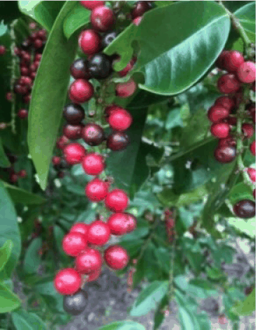
Antidesma thwaitesianum Müll. Arg., commonly known as Mao or Ma-Mao or Mak-Mao in Thai (Fig. 1), belongs to the family Stilaginaceae and the genus Antidesma (Jorjong et al., 2015). Mao is a medium-sized evergreen tree with oval to oblong leaves with pointed tips and smooth surfaces. The unisexual, small, and yellowish-white colored flowers bloom in clusters at the tips of branches. The fruits are round, grow in bunches, and exhibit a light-white color when unripe, transitioning to red or dark-purple color depending on the level of ripeness. They have a sweet and sour flavor (Hoffmann, 1999). Ripe Mao fruits have gained popularity, and are processed into health products such as Mao juice and wine, owing to their phytochemical components that possess antioxidant properties. In 2019, the total production of Mao in Thailand reached 491 tons (Department of Agricultural Extension, 2020), with the average cultivation cost for farmers amounting to 61,906 baht/hectare (Sriphadet & Phukna, 2017). During processing, approximately 30%–40% of Mao remains as waste and by-products (Butkhup & Samappito, 2008). Mao residue has been utilized in agriculture due to its beneficial compounds, including phenolics and flavonoids (Hansakul et al., 2015; Kittipongpittaya et al., 2021), catechin, procyanidin B1, procyanidin B2 (Butkhup & Samappito, 2008), polyphenols (97.32–130 mg/g gallic acid equivalents), proanthocyanidin (Puangpronpitag et al., 2008), tartaric acid (0.16–0.22 g/100 g), malic acid (0.03–0.05 g/100 g) and citric acid (0.15–0.43 g/100 g) (Lokaewmanee & Sansupha, 2015). Furthermore, Mao extract has been shown to alleviate hypertension and oxidative stress in nitric oxide deficient rats (Kukongviriyapan et al., 2013). Additionally, Mao extract has been reported to exhibit antibacterial activity against Streptococcus, including Streptococcus constellatus, Streptococcussalivarius, and Streptococcusmitis (Sotthisawad & Insin, 2012), and has demonstrated effectiveness in reducing the bacterial load of Streptococcus spp. (Kamolrat et al., 2021). The objective of this study is to investigate the effects of Mao residue extract on A. hydrophila and S. agalactiae, two pathogenic bacteria responsible for diseases in Nile tilapia, as an alternative to reducing the use of antibiotics in fish farming.
Materials and Methods
This study was conducted in strict accordance with the guidelines for the use of animals regulated by the Institute of Animals for Scientific Purposes Development (IAD), Thailand. Fish handling and all experimental protocols were approved by the ethics committee at Rajamangala University of Technology Isan (Approval number: 9/2020).
Mao residue was obtained from the production of Mao juice at the Agro-tourism Inpang center, Sakon Nakhon, Thailand. The Mao marc was dried using a hot air oven at a 60°C for 72 hours and then ground into a fine powder. The dried ground Mao marc was extracted using varying polarity solvents: distilled water (highly polar solvent), 50% ethanol (hydro-ethanolic mixture), or 95% ethanol (less polar) at a ratio of 1:3 g/ml. The extraction process involved soaking the material in the respective solvents for 72 hours at room temperature. The solvents were removed using a rotary evaporator. The resulting crude extracts were weighed and stored in an amber glass bottle at 4°C for use throughout the experiment. The percentage yield of the extract was calculated as follows: % Yield = Weight of the extract (g) × 100/Weight of initial sample before extraction (g).
Qualitative phytochemical analysis of the crude extracts was conducted using different solvents. Alkaloids were identified using Wagner’s test methods, where the formation of brownish-red precipitate indicated a positive result (Fig. 2). Tannins were detected by adding ferric chloride to the extract; the appearance of brown-dark green coloration signified a positive result (Fig. 2). Saponins were tested using foam test. The extracted solvent was mixed with distilled water in a graduated cylinder and shaken for about 10–15 minutes, where the appearance of about 1 cm thick layer of foam indicated a positive result. Flavonoids were detected using a 10% lead acetate solution, with the formation of yellow color indicating a positive result (Fig. 2). The extract solution was hydrolyzed with dilute sulfuric acid and subsequently extracted with benzene, with further addition of ammonia producing a rose-pink color and indicating the presence of anthraquinones. Glycosides were detected using sodium nitro-prusside solution, with the formation of pink-red color indicating a positive result (Fig. 2). Steroids were identified by mixing equal volumes of chloroform and sulfuric acid. The appearance of two separate layers (an upper red layer and a yellow-green fluorescent sulfuric acid layer) indicated the presence of steroid in test sample (Kumar et al., 2013).
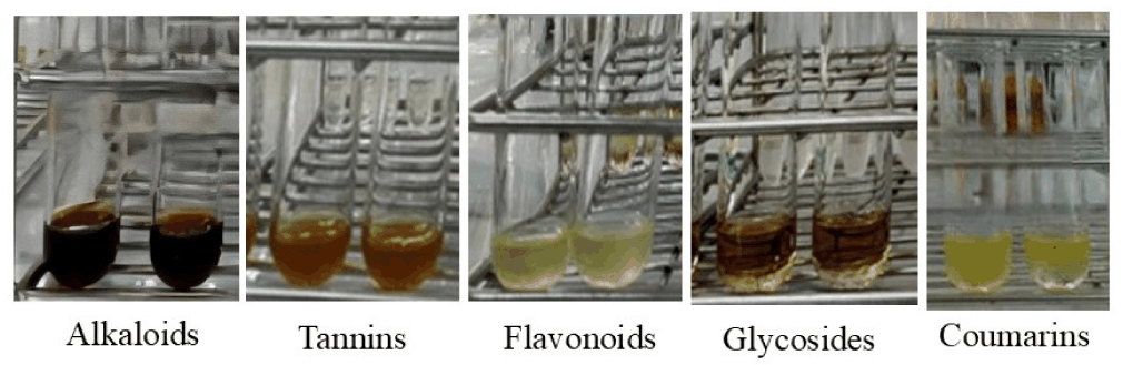
Phlobatannins were tested using the precipitate test. The extracted solvent was mixed with hydrochloric acid and the solution subsequently heated, with a red precipitate indicating a positive result. Chloroform and sulfuric acid were used to detect terpenoids, with a deep red coloration indicating a positive result. Sodium hydroxide solution was used to test the presence of coumarins; a formation of yellow coloration showed a positive result (Fig. 2) (Yadav et al., 2014).
The bacterial strains (A. hydrophila and S. agalactiae serotype Ia) used in this experiment were obtained from the School of Agricultural Technology and Food Industry, Walailak University, Thailand. The bacteria were grown on tryptic soy agar (TSA) media and incubated at 37°C for 24 hours. Following incubation, the bacterial cultures were diluted with 0.85% saline solution to prepare bacterial suspensions. The density was measured using a spectrophotometer at OD600nm value of 1 to the density of A. hydrophila of 1 × 10⁸ CFU/ml (Crumlish et al., 2010). A wavelength of OD540nm value of 0.15 was used for S. agalactiae to the density of 1 × 10⁸ CFU/ml (Nithikulworawong, 2012).
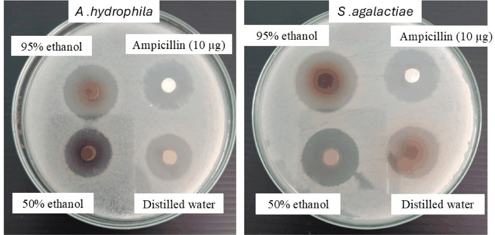
The antibacterial was assessed using the disc-assay method. The prepared bacterial inoculum was swabbed onto TSA plates. Extract from each solvent was dropped onto a 6 mm diameter paper disc at a concentration of 1,000 mg/ml at a volume of 30 µl (Dilbato Dinbiso et al., 2022) and then placed on the surface of the medium. The plates were then incubated at 37°C for 24 hours. Ampicillin (10 µg) was used as positive control. The antibacterial activity was evaluated by measuring the diameter of the inhibition zone in millimeter (mm).
Minimal inhibitory concentration (MIC) and minimal bactericidal concentration (MBC) assays were conducted using a broth dilution method modified from the Clinical and Laboratory Standards Institute (CLSI) M07 (CLSI, 2012) guidelines. The modifications include the use of tryptic soy broth (TSB) as the culture medium and adjustment of bacterial inoculum to a density of 1 × 10⁸ CFU/ml for both A. hydrophila and S. agalactiae. The antimicrobial efficacy of Mao extract by evaluating the visible growth of microorganisms in the agar broth. The high initial concentration (500 mg/ml) was chosen due to plant extracts often contain complex mixtures of phytochemicals with variable solubility and antimicrobial efficacy, which are generally lower than purified antibiotics. The extract with the highest inhibition zone was selected and subjected to a two-fold serial dilution to obtain concentrations of 500, 250, 125, 62.5, 31.25, 15.62, 7.81, 3.90, 1.95, 0.97, and 0.49 mg/ml. Then, 0.01 ml of the prepared A. hydrophila and S. agalactiae bacterial suspensions were pipetted into 1.5 ml of TSB culture medium and incubated at 37°C for 24 hours. In the control group, no extract was added. The MIC was defined as the lowest concentration that showed no visible bacterial growth compared with the control. All the MIC tubes with no visible growth were plated out on TSA agar and incubated at 37°C for 24 hours. The MBC was defined as the lowest concentration at which no bacterial colonies were observed on TSA agar.
Nile tilapia fries with an average weight of 3.38 ± 0.04 g were stocked in cement tanks at the Division of Fisheries, Rajamangala University of Technology Isan, Sakon Nakhon campus. Fish were fed a commercial diet containing 32% protein and 4% lipid (Charoen Pokphand Foods, Thailand) twice a day at a rate of 5% of body weight, once in the morning and once evening, for 7 days. Prior to feeding, the feed was supplemented with 500 mg/ml of Mao extract, extracted using a 95% ethanol solvent, coated with fish oil, and allowed to air dry indoors. The experiment was conducted using 300 L fiber tanks, with a stocking density of 60 fish per tank. A completely randomized design (CRD) was employed, with four treatment groups in triplicate as follows: Treatment 1 (control): fish fed the standard commercial diet; Treatment 2: fish fed the commercial diet supplemented with 0.1% Mao extract; Treatment 3: fish fed the commercial diet supplemented with 0.5% Mao extract; Treatment 4: fish fed the commercial diet supplemented with 1.0% Mao extract. Fish were fed twice a day (08:00 h and 17:00 h) for 8 weeks. The extract-coated diet was freshly prepared for each feed. Throughout the experiment, fish weight, body length and survival rates were measured every 2 weeks. Water quality parameters were monitored throughout the trial to ensure they remained within acceptable ranges for fish culture (temperature of 29 ± 1°C, pH of 7.8 ± 0.5, dissolved oxygen of 6.5 ± 0.5 mg/l, and ammonia nitrogen below 0.05 mg/l).
At the end of the experiment, the total number of fish, as well as their individual body length and weight from each tank were measured to calculate the survival and growth performance. Growth parameters for each replicate were using the following formulas: Weight gain (WG) = 100 × (Final body weight – Initial body weight)/Initial body weight; Average daily gain (ADG) = (Average final weight – Average initial weight)/Feeding period; Food conversion ratio (FCR) = Dry feed intake/Wet WG; Survival rate = 100 × (Final fish number)/(Initial fish number).
After 8 weeks, Nile tilapia was tested for antibacterial resistance. All treatment groups were intraperitoneally injected with A. hydrophila and S. agalactiae serotype Ia at a concentration of 108 CFU/ml in a volume of 0.1 ml. This concentration was selected based on previous report of LD50 values at 108 CFU/ml for A. hydrophila (Mzula et al., 2020) and 108 CFU/ml for S. agalactiae (Owatari et al., 2022). The control groups were divided into two: Group 1 was injected with 0.85% normal saline (negative control) and Group 2 was injected with the same bacterial concentration (positive control). The mortality of the fish was recorded daily for 21 days. The relative percent survival (RPS) for each treatment group was calculated using the following formula: RPS (%) = 100 × [1 – (% Mortality in treatment group/% Mortality in control group)] (Amend, 1981). Fish were not euthanized prior to bacterial challenge, as the objective was to assess disease resistance following exposure. Humane endpoints were strictly followed: fish exhibiting severe clinical signs (e.g., loss of equilibrium, unresponsiveness, or dying condition) were immediately euthanized with an overdose of MS-222 (200 mg/l). At the termination of the trial, all surviving fish were humanely euthanized.
Results
The characteristics and yield of Mao residue extracts obtained using different solvents are shown in Table 1 and Fig. 3. The 95% ethanol extract provided the highest yield of crude extracts compared to other solvents.
The phytochemical analysis of Mao residue extract is shown in Table 2. The extraction with various solvents revealed the presence of various secondary metabolites, including flavonoids, coumarins, alkaloids, saponins, tannins, and glycosides. The 95% ethanol extract exhibited more active compounds compared to other solvents (Table 3).
The MIC test for Mao residue extracts revealed that aqueous, 50% ethanol, and 95% ethanol extracts each inhibited the growth of A. hydrophila at a concentration of 62.5 mg/ml. For S. agalactiae, the 95% ethanol extract was the most effective, also exhibiting an MIC of 62.5 mg/ml (Table 4). In the MBC assay, for A. hydrophila, all three extract types exhibited bactericidal activity at 250 mg/ml. For S. agalactiae, only the 95% ethanol extract demonstrated bactericidal activity at 500 mg/ml.
| A. hydrophila (gram-negative) | S. agalactiae (gram-positive) | ||||
|---|---|---|---|---|---|
| Distilled water | 50% ethanol | 95% ethanol | Distilled water | 50% ethanol | 95% ethanol |
| 62.5 | 62.5 | 62.5 | 250 | 250 | 62.5 |
Based on these results, the 95% ethanol extract of Mao residue was selected for further testing of its inhibitory efficacy against A. hydrophila and S. agalactiae in Nile tilapia fry, as it demonstrated the highest antibacterial activity against both bacterial species.
After the 8-week feeding trial, Nile tilapia fed with a commercial diet supplemented with varying concentrations of Mao extract showed no statistically significant differences (p > 0.05) in initial weight, final weight, WG, ADG, feed conversion ratio, or survival rate across all experimental groups (Table 5).
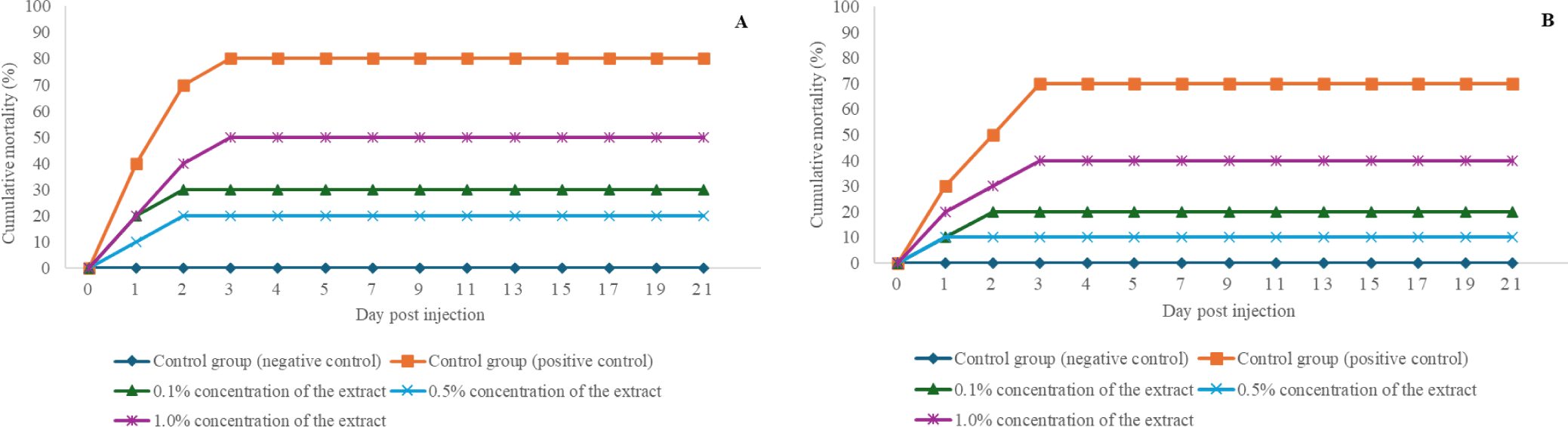
The antibacterial resistance study in Nile tilapia showed that fish fed a commercial diet supplemented with 0.5% Mao extract exhibited the highest resistance to A. hydrophila and S. agalactiae. The A. hydrophila challenge revealed a mortality rate of 20%, with a relative survival of 75% (Table 6), resulting in 20% cumulative mortality (Fig. 4A). In the S. agalactiae challenge, the mortality rate was 10% (Table 6), with a relative survival of 85.71%, resulting in 10% cumulative mortality (Fig. 4B).
Discussion
Over the past decade, herbs and various parts of plants have been increasingly incorporated into aquatic animal diets due to their pharmacologically active compounds. Many studies have identified A. thwaitesianumfruit as a rich source of polyphenols and proanthocyanidins (Puangpronpitag et al., 2008) as well as total phenolics and flavonoids (Hansakul et al., 2015; Kittipongpittaya et al., 2021). Several pharmacological effects of A. thwaitesianum have been reported, including anti-alpha amylase, anti-alpha glucosidase, anticancer, antitumor, anti-apoptotic, anti-inflammatory, antimicrobial, anti-viral and antioxidant activities (Mahomoodally et al., 2012; Nguyen-Ngoc et al., 2024; Puangpronpitag et al., 2011).
The Mao residue extract exhibited antibacterial efficacy against A. hydrophila and S. agalactiae with a MIC of 62.5 mg/ml. This study demonstrated that Mao extract showed bactericidal activity against both gram-positive and gram-negative. However, other studies showed lower antimicrobial activity of Mao against multiple bacterial strains. Tinchan et al. (2022) found Mao juice had antibacterial activity against three gram-positive (Bacillus cereus, Streptococcus aureus, and Listeria monocytogenes) and three gram-negative (Salmonella typhimurium, Pseudomonas aeruginosa, and Escherichia coli). L. monocytogenes was the most sensitive to Mao juice with MIC of 25 mg/ml, while a MIC of 50 mg/ml for B. cereus, S. aureus, S. typhimurium, and E. Coli. Sriphadet & Srisopa (2021) also showed antimicrobial action of Mao against S. typhimurium, B. cereus, and E. Coli, with MIC of 25 mg/ml. Dechayont et al. (2012) found dry marc extract of Mao exhibited antimicrobial activity against S. aureus, with MIC of 2.5 mg/ml. Pongnaratorn et al. (2017) also reported Mao fruit extracts had inhibitory effects on S. aureus, with MIC of 0.1 mg/ml and inhibitory effects on Streptococcus mutans and Streptococcus pyogenes with both MIC of 0.05 mg/ml.
The action of Mao extracts on against A. hydrophila and S. agalactiae shows moderate antimicrobial activity. The extract’s efficacy against A. hydrophila compared to other phytochemical, this concentration is similar to turmeric (Curcuma longa) extract (62.5 mg/ml; Sincharoenpokai et al., 2009). However, the concentration required for Mao extract was higher than the reported MIC value for various other plant extracts, including Syzygium aromaticum (12.5 mg/ml; Najiah et al., 2011), Bauhinia sirindhorniae (6.25 mg/ml; Nithikulworawong, 2012), Cassia fistula (1.5 mg/ml; Borisutpeth et al., 2005), as well as Bidens pilosa (0.625 mg/ml; Son et al., 2022). Conversely, Mao extract was more effective than garlic (Allium sativum) extract, which required 100 mg/ml to inhibit similar activity (Artawinata et al., 2025).
In the case of S. agalactiae, the MIC of Mao extract was the same as Azadirachta indica and Olea europaea extracts (both 62.5 mg/ml; Abdallah et al., 2024), as shown by Wei & Musa (2008), who obtained MIC of 62.5 mg/ml using garlic extract. However, when compared to other plant extracts, it weaker than Psidium guajava (25 mg/ml; Wattanuruk & Detraksa, 2023), Excoecaria agallocha (6.25 mg/ml; Abdul Razak et al., 2019), C. longa (6.25 mg/ml; Pisuttharachai et al., 2020), Combretum quadrangulare (3.125 mg/ml; Tran et al., 2021), Punica granatum (2.5 mg/ml; Nakhubon et al., 2022), Calyptranthes clusifolia (1.5 mg/ml; Castro et al., 2008), as well as the essential oils of Mentha piperita (1.25 mg/ml; Majolo et al., 2018).
Qualitative screening has revealed the presence of various bioactive compounds, including flavonoids, coumarins, alkaloids, saponins, tannins, and glycosides, all of which are recognized for their antibacterial properties.
The antibacterial activity observed in this study may be attributed to the presence of several bioactive phytochemicals identified in Mao residue extracts. Flavonoids have been reported to inhibit nucleic acid synthesis and damage bacterial cell walls (Cushnie & Lamb, 2011). Tannins can form complexes with microbial proteins and enzymes, thereby impairing bacterial growth (Scalbert, 1991). Saponins have surfactant-like effects that increase membrane permeability and cause cell lysis (Francis et al., 2002), while alkaloids are known to disrupt bacterial metabolism (Cushnie et al., 2014). In addition, Puangpronpitag et al. (2008) also reported that the extract of Mao marc contained a high amount of polyphenolic compound. The phenolic compounds, particularly catechins, gallic acid derivatives, and anthocyanins, could disrupt the bacteria cell wall, which protects the bacteria from toxic compounds (Lizardo et al., 2015). The combined presence of these compounds probably contributed to the inhibitory effects against A. hydrophila and S. agalactiae observed in this study.
No significant differences were observed in final weight, WG, ADG, feed conversion ratio, or survival rate among Nile tilapia fed different concentrations of Mao extract. This study is the first to investigate the use of Mao extract as a dietary herbal supplement for Nile tilapia, with the findings indicating that Mao extract supplementation does not affect growth performance parameters.
This study demonstrated that dietary supplementation with Mao extract increased the survival rate of Nile tilapia following challenge with A. hydrophila and S. agalactiae. Compared to the fish fed without the Mao extract, the 0.5% Mao extract showed tolerance to both bacterial challenges, leading to a higher survival rate and with RPS against A. hydrophila and S. agalactiae of 75.0% and 85.7%, respectively. The protective efficacy of Mao extract was comparable to several well-known phytogenic agents, such as olive leaf extract (0.1% feed inclusion against A. hydrophila; 90% survival; Assar et al., 2023) and allspice powder (10 g/kg feed against S. agalactiae; 80% survival; Yılmaz & Ergün, 2014). Similarly, the high RPS value (78.9% and 87.4%) was detected in guava and star gooseberry leaf extracts in the resistance of Nile tilapia against A. hydrophila (Kamble et al., 2024).
One interesting result of this study is that the dose-response relationship is non-linear. Where 0.5% Mao extract supplementation provided better RPS value than those at 1% concentration. This trend contradicts the traditional dose-dependent expectations but aligns with several reported phytogenic interventions in aquaculture. Kamolrat et al. (2021) observed that 20% of Mao juice concentration mixed with feed powder resulted in a lower cumulative mortality against S. agalactiae compared to 40% and 60% concentrations. Similarly, Yılmaz et al. (2022) reported that fish fed 2% black mulberry syrup had a higher RPS than 3% supplementation, and Baba et al. (2016) found that the highest resistance of A. hydrophila occurred at 10 g/kg oat extract, rather than higher concentrations. This was also supported by Acar et al. (2015), where 0.1% citrus essential oil proved more protective in Nile tilapia compared to higher dose in Streptococcus iniae. Previous studies have reported that high concentrations of herbal supplements can have adverse effects. For instance, Zemheri-Navruz et al. (2019) observed that high concentration of olive leaf extract suppressed immune function in common carp, while Baba et al. (2018) reported that allergic reactions as side effects of herbals due to their constituents in excessive doses. Biller & Takahashi (2018) suggested that high antioxidant levels might reduce oxidative stress, subsequently weakening the innate immune response in fish and increasing susceptibility to disease.
This result indicated that Mao extract had a positive effect on survival rate of Nile tilapia, and this could be due to the cooperative effects of active compounds in the extract. However, this work primarily assessed the in vitro antibacterial properties of Mao extracts. Although some medicinal herbs are known to enhance fish innate immunity, our study did not directly measure immune responses. Therefore, claims regarding immunomodulatory effects cannot be drawn from the present findings, and further in vivo investigations are recommended.
Conclusion
The findings of this study indicate that Mao extract inhibitory effects against A. hydrophila and S. agalactiae. Fish fed a commercial diet supplemented with 0.5% Mao extract demonstrated the highest resistance to bacterial infections. These findings suggest that Mao extract shows potential as a feed additive to enhance disease resistance against A. hydrophila and S. agalactiae infections. Nevertheless, the application of herbal medicines in the aquaculture industry warrants further investigation.