Introduction
Nanotechnology, a well-established field of research since the last century, has the potential to impact the livestock and agriculture industries worldwide. Nanoparticles, nanosensors, microfluidics, and bioanalytical devices are examples of nanotechnology tools employed in these sectors (Gelaye, 2024). Numerous macro ingredients are commonly used as additives in animal feed preparation. Additionally, nanoparticles are gaining attention as feed additives due to offering several benefits, like enhanced bioavailability, reduced dosage needs, and effective component interaction. These additives help improve nutrient intake, absorption, and utilization while also promoting growth and supporting physiological functions such as immune enhancement, stress management, and reproductive health (Onuegbu et al., 2018). Research on trace minerals in nanoparticle form has shown promise as an alternative for meeting mineral needs in livestock and poultry feed. Their minimal dosage requirements make them an attractive substitute for antibiotics, encouraging extended use, reducing traces of antibiotics in animal-derived products, lowering environmental pollution, and ensuring clean and uncontaminated animal products (Schmidt, 2009). In aquaculture, researchers have investigated the use of different metallic and nonmetallic nanomaterials in the diets of different fish species, and results showed improvements in the rate of growth, feed intake, immune response, and resistance to diseases (Dube, 2024; Rakhi et al., 2022).
Besides trace minerals, unconventional metals are becoming a focus of research as feed ingredients. Studies have reported on the effects of chromium, titanium oxide, and gold nanoparticles in fish feed, examining their impact on growth performance, mineral absorption, immunostimulation, and toxicity (Ahmad et al., 2023; Shahariar et al., 2024). However, dietary studies on silver nanoparticles (AgNPs) in fish are still limited. AgNPs possess impressive optical, morphological, and chemical properties (Abbas et al., 2024). Silver ions are released more efficiently with a greater surface area, suppressing bacterial growth and boosting their antimicrobial capabilities (More et al., 2023). Zaoui et al. (2024) reported that AgNPs supplementation (10 mg kg–1 kaolin) improves the feed conversion ratio in broiler chickens. AgNPs at 20 parts per billion were reported to positively impact growth and metalloprotease activity in the gut of zebrafish (Sarkar et al., 2015). Popoola et al. (2023) demonstrated that dietary inclusion of AgNP at 15 µg/Kg significantly enhanced growth, overall health, and protective immune against Aeromonas hydrophila in Labeo rohita. Similarly, Kumar et al. (2018) reported that AgNPs supplementation at 0.5 mg kg–1 improved growth performance, immune responses, survival rates, and reduced stress biomarkers such as HSP70, cortisol, and blood glucose levels in various tissues of Channa striatus. In poultry, silver-doped silica nanoparticles were identified as promising and safe nano-growth promoters when administered at 4 mg kg–1 in broiler diets (Dosoky et al., 2021).
However, data on the impacts of biosynthesized AgNPs on fish growth and hematological parameters at higher dietary doses are limited. AgNPs synthesized using plant extracts are rich in phytochemicals that help reduce Ag+ to form the nanoparticles (Ahmed et al., 2016). In this study, mango leaf (Mangifera indica) extract, which is rich in phytochemicals, namely phenolic compounds, flavonoids, apigenin, phenols, benzophenones, carbohydrates, terpenoids, and quinones, was selected to biosynthesize AgNPs (Kumar et al., 2021). The striped dwarf catfish (Mystus vittatus) was selected as a model fish species to evaluate nanoparticle-based dietary interventions in aquaculture. As yolk-absorbed hatchlings are introduced into rearing ponds, they face the transition from planktonic feed to other natural food sources. They must adapt to a new environment, moving from enclosed hatcheries to open nursery ponds. Ensuring adequate nutrition during this stage in the nursery ponds to build strong immunity against diseases can help achieve optimal survival and growth rates (Mou et al., 2018). Incorporating biosynthesized AgNPs into the feed of striped dwarf catfish holds significant promise in this regard. Therefore, this study aimed to evaluate the effects of biosynthesized AgNPs as dietary supplements in M. vittatus, focusing on growth performance, body composition, survival rate, and hematological parameters.
Materials and Methods
Silver nitrate (analytical grade AgNO3) was obtained from MP Biomedicals (Illkirch, France). Deionized water was used throughout the experiment. Fresh mango leaves (M. indica) collected from the research area were first properly washed, sun-dried, and ground into powder. Ten grams of ground leaf powder were mixed with 100 mL distilled water and heated at 80°C under vigorous stirring for 20 min. After cooling, the mixture was filtered (Whatman Grade 1) to prepare the extract. Then, 10 mL of the extract were added to 50 mL of 1.0 mM AgNO3 solution. The reaction mixture was stirred at 220 rpm on a hot-plate magnetic stirrer for 20 min until the color changed from reddish-brown to dark brown. The suspension was centrifuged at 18,200 ×g for 15 min (Velocity 18R). The precipitate was washed with distilled water and ethanol and then freeze-dried under vacuum. The AgNP powder was characterized by UV-Vis spectral analysis, X-ray diffraction (XRD), Fourier transform infrared (FTIR) spectroscopy, and transmission electron microscopy (TEM) with energy-dispersive X-ray spectroscopy (EDS), following Shahariar et al. (2024).
The experimental feed included AgNPs at 0 mg kg–1 (control), 10 mg kg–1 (T10), 30 mg kg–1 (T30), and 50 mg kg–1 (T50) in a commercial pellet feed (Mega Feed, Spectra Hexa Feed Limited, Jashore, Bangladesh). According to the packaging, pellet diameter was 0.6 mm with 37% protein, 8% lipid, and < 16% ash. For each treatment, 600 g of diet was prepared. Along with the control diet, the pellets were first ground to ensure even blending to make all the experimental diets. An ultrasonic bath (Elmasonic P, Elma, Germany) was used for 10 min to disperse AgNPs in distilled water before homogeneous mixing with the ground feed. The mixture was thoroughly combined with the ground feed, re-pelleted, and sun-dried for 6 h. The prepared diets were stored refrigerated until use.
Fish were collected from a hatchery one week before the experiment and acclimatized to tanks at the experimental site. The experiment was conducted in rectangular glass aquariums with 70 L water-holding capacity (Afrin et al., 2023). Three replicates were used for each treatment. Initially, 60 fish were weighed and stocked in each aquarium. Every 15 days, 20 fish from each aquarium were weighed to assess biomass. Fish were fed the experimental diets for 75 days, three times daily (morning, afternoon, and late evening) to apparent satiation.
Temperature, ammonia, pH, dissolved oxygen (DO), and total dissolved solids (TDS) in control, T10, T30, and T50 treatments were measured at 9:00 h at five-day intervals.
Growth parameters—weight gain percentage (WG%), specific growth rate (SGR; % day–¹), protein efficiency ratio (PER), feed conversion ratio (FCR), condition factor (CF), and survival rate (SR%)—were calculated following Hossain et al. (2022).
Blood was collected in ethylenediaminetetraacetic acid (EDTA) tubes by puncturing the caudal peduncle. For each replicate, blood samples from 10 fish per aquarium were collected. Hemoglobin (Hb), red blood cell (RBC) count, white blood cell (WBC) count, mean corpuscular volume (MCV), mean corpuscular hemoglobin (MCH), and mean corpuscular hemoglobin concentration (MCHC) were determined using an automated hematology analyzer (Lima et al., 2024).
Results
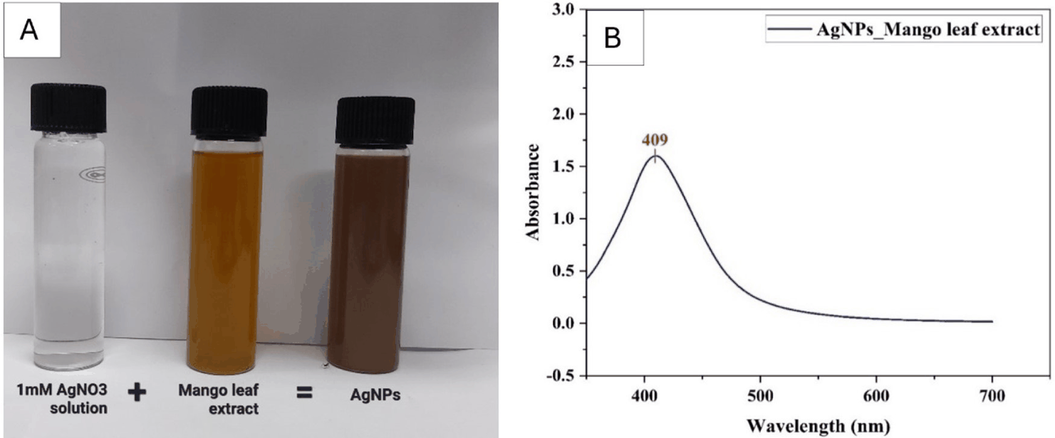
A color change from reddish-brown to dark brown (Fig. 1A) indicated successful AgNP synthesis. UV-Vis spectrum analysis showed a peak at 409 nm (Fig. 1B), consistent with the surface plasmon resonance of AgNPs.
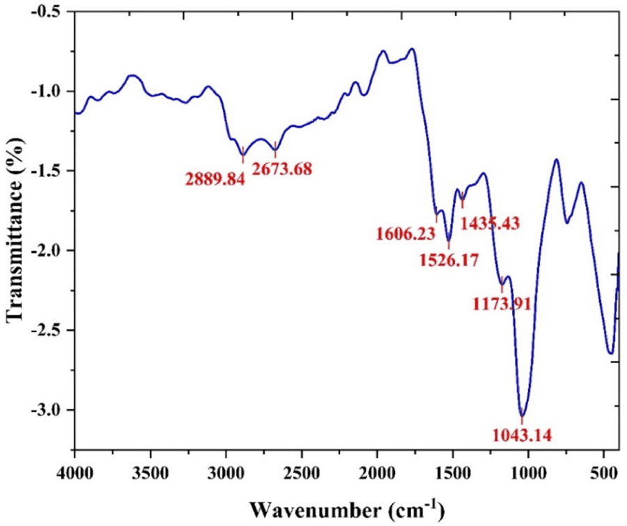
FTIR analysis was used to identify possible capping biomolecules (Table 1). Seven bands were observed at ~2,889.84, 2,673.68, 1,606.23, 1,526.17, 1,435.43, 1,173.91, and 1,043.14 cm–¹ (Fig. 2).
| Bands (cm–1) | Possible functional group | References |
|---|---|---|
| 2,889.84 | C-H stretching of aromatic compound | Jyoti et al. (2016) |
| 2,673.68 | O-H carboxylic acids | Subarani et al. (2013) |
| 1,606.23 | N-H bend primary amines | Mahitha et al. (2011) |
| 1,526.17 | C-H plane deformation vibrations of methyl, methylene, and methoxy groups | Bankar et al. (2010) |
| 1,435.43 | C-N stretching of the aromatic amine group | Mahitha et al. (2011) |
| 1,173.91 | C-N stretching of amines | Jyoti et al. (2016) |
| 1,043.14 | C-N aliphatic stretch | Mahitha et al. (2011) |
XRD analysis assessed the purity and composition of the products yielded through biosynthesis utilizing M. indica extract. Fig. 3 shows the XRD pattern recorded over 10°–80° 2θ with a step size of 0.02°.
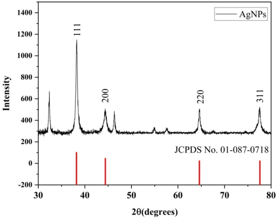
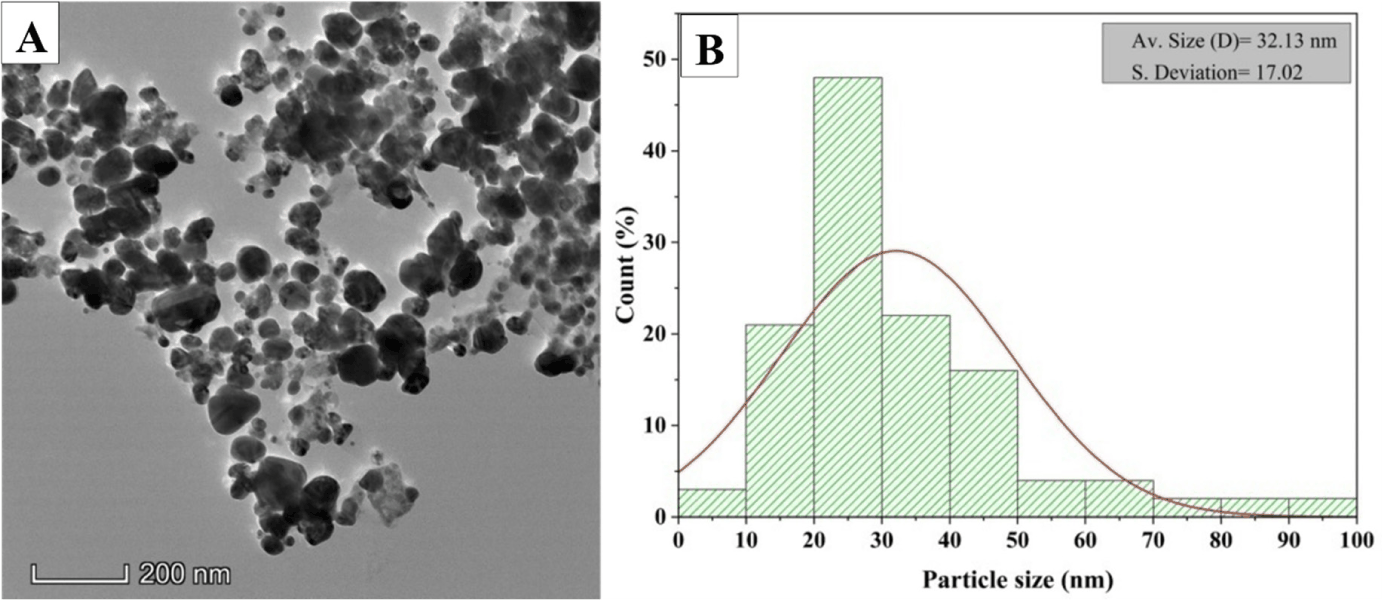
XRD showed Bragg peaks at 38.2°, 44.35°, 64.62° and 77.46°, corresponding to the (111), (200), (220), and (311) planes for face-centered cubic silver (JCPDS [Joint Committee on Powder Diffraction Standards] card no. 01-087-0718). The crystallite size was ~23.52 nm based on the full width at half maximum of the (111) peak (Debye–Scherrer). TEM showed predominantly spherical polydisperse particles with an average size of 32.13 nm and diameters ranging from 5 to 93.6 nm (Fig. 4). EDS confirmed the presence of silver in the nanoparticles (Table 2 and Fig. 5).
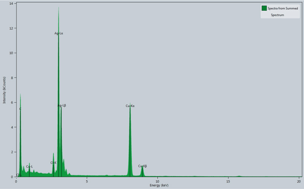
| Element | Weight | Atomic |
|---|---|---|
| Silver (Ag) | 59.79 | 27.90 |
| Copper (Cu) | 26.71 | 21.16 |
| Carbon (C) | 11.47 | 48.06 |
| Chlorine (Cl) | 2.02 | 2.87 |
Table 3 summarizes physicochemical data (temperature, dissolved oxygen, pH, ammonia, and total dissolved solids) measured during the experimental period.
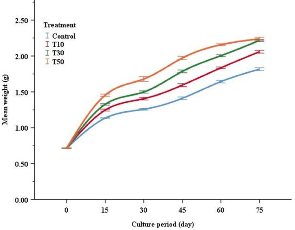
Fig. 6 illustrates changes in average weight of M. vittatus over the 75-day period. Weight gain (%) increased with dietary AgNPs up to 50 mg kg–1 and differed from the control (p < 0.05) (Table 4). The T30 treatment (30 mg kg–1) showed higher WG%, SGR(% day–1), SR%, and improved FCR and PER relative to the control (p < 0.05).
Significant differences (p < 0.05) are denoted by different superscript letters within the same row.
Hematological parameters (RBC, Hb, WBC, hematocrit [HCT, %] and plateletcrit [PCT, %], MCV, MCH, and MCHC)are shown in Table 5. T30 exhibited higher RBC count, Hb, and MCH, whereas WBC did not differ significantly among treatments (p > 0.05).
Significant differences (p < 0.05) are denoted by different superscript letters within the same row.
Discussion
The AgNP synthesis was indicated by a color change and further confirmed by UV–Vis peak at 409 nm. Similar to our study, Hai et al. (2022) recorded peaks from 380 to 450 nm. The particle size range was much larger (5 to 220 nm; average 52.8 nm) when biosynthesized using mango leaf extract at room temperature compared to this study (5 to 93.6 nm; average 32.13 nm), though the shape was similar (Alqahtani et al., 2022). Although DO and TDS differed significantly among treatments, all water quality parameters (temperature, DO, pH, ammonia, and TDS) remained within the optimal ranges (Ghafarifarsani et al., 2024). We also acknowledge that water quality parameters were measured at fixed morning intervals during the trial. While this ensured consistency across treatments, more frequent or continuous monitoring may better capture diurnal variations and should be considered in future experimental designs. Moreover, through visual inspection, it was evident that the experimental fish actively took the feed, reducing the chance of AgNP leaching into the water and affecting water-quality parameters.
In this study, growth metrics demonstrated an upward trend with the addition of AgNPs, ranging from 0 mg kg–1 to 50 mg kg–1. Significant differences in WG% and SGR (% day–1) were observed in the 10 mg kg–1 (T10), 30 mg kg–1 (T30), and 50 mg kg–1 (T50) AgNPs diets compared to the 0 mg kg–1 (control) AgNPs diet (p < 0.05). Significantly better growth performance was observed in T30 compared to other treatments. Although growth data were collected at multiple time points during the 75-day trial, only the final values were statistically analyzed using one-way ANOVA to compare treatment effects. This decision was based on the design of the study and aligns with previous nanoparticle feed trials (e.g., Shahariar et al., 2024). We acknowledge that repeated-measures or mixed-effects models may provide greater statistical resolution and plan to adopt such approaches in future studies. Although post-formulation analysis of silver content in the prepared diets was not performed due to equipment limitations, uniform dispersion of nanoparticles was ensured through ultrasonic and manual mixing techniques. Future studies will include silver quantification using Inductively Coupled Plasma Optical Emission Spectroscopy (ICP-OES) or similar methods to confirm dose accuracy. In our study, AgNPs dietary incorporation levels (0, 10, 30, and 50 mg kg–1) were selected based on an exploratory approach to assess their effects on fish growth performance. This suggests that at this concentration, AgNPs may play a role in improving metabolic efficiency and nutrient utilization. While previous studies have primarily examined lower AgNPs concentrations in waterborne exposure or feed, our results demonstrated that fish fed with 30 mg kg–1 AgNPs-incorporated feed exhibited the highest growth, with no visible signs of abnormal behavior, altered movement, or reduced feeding activity. Popoola et al. (2023) suggested that dietary inclusion of AgNPs at 10 and 15 µgKg–1 feed enhances growth, health, and protective immune response against A.hydrophila. Aqueous exposure to AgNPs (6 and 9 μg/L) synthesized by abalone viscera hydrolysates promotes the growth, immunity and gut health of Danio rerio and induced catalase and superoxide dismutase activities and increased glutathione (GSH) content in the livers and upregulated the expression of immune related genes (Ni et al., 2022). Shyamala & Maheswari (2024) suggested 10 μg/kg Pleurotus ostreatus mediated AgNPs as a beneficial dietary supplement for improved hematological, immunological response and disease resistance in tilapia against A. hydrophila. Sarkar et al. (2015) reported that the inclusion of AgNPs in the diet of D. rerio at the rate of 20 parts per billion (ppb) exhibited the highest WG and SGR, followed by 40 ppb compared with other treatments. Zaoui et al. (2024) also reported that AgNP supplementation improved the feed conversion ratio in broiler chickens. Also, 12 to 15 percent better growth and FCR (3.62 to 7.38 percent) were observed in broilers supplemented with 2 to 10 mg nano-Ag/kg of feed compared to the control (Elkloub et al., 2015). Previously published results showed that the inclusion of AgNPs in poultry feeds is inconsistent. Some studies reported zero significant responses to growth performance after the addition of AgNPs (Ahmadi et al., 2013). Applying AgNPs to the egg (in ovo) can upregulate the expression of fibroblast growth factor and vascular endothelial growth factor, potentially stimulating the proliferation and differentiation of satellite cells (Sawosz et al., 2012). Moreover, it was reported that the expression of genes responsible for muscle development during embryogenesis in chicken embryos was affected by injected AgNPs (Pineda et al., 2012; Sawosz et al., 2012). An in vitro study showed that silver nanoparticle treatments significantly reduced the coliforms in pig ileal lysate. This reduction was enhanced when the doses of AgNPs were proportionally increased (Fondevila et al., 2009). The fish are likely less vulnerable to pathogenic problems in the stomach due to the antibacterial action of the AgNPs, which improves digestive processes. Similarly, trace minerals like nano iron and nano zinc oxide supplemented diets also improved the growth of the fish (Nirmalkar et al., 2023). It is important to acknowledge that cellular-level toxicity assessments were not conducted in this study. However, given that no external morphological or behavioral abnormalities were observed, it is reasonable to infer that these doses were well-tolerated within the experimental period. Previous research has highlighted dose-dependent effects of AgNPs, where beneficial impacts on immunity and growth were observed at optimal levels, but higher concentrations led to oxidative stress and tissue damage. While our study did not extend to histological examination, the control and AgNPs-treated fish exhibited comparable survival rates and growth performance, reinforcing the potential of 30 mg kg–1 as an effective dose. Future research should include cellular and molecular-level analyses to validate the long-term safety and physiological effects of AgNPs dietary incorporation in fish. Specifically, assessments such as oxidative stress markers, cytokine profiling, and histopathological evaluation of tissues will be important to validate long-term safety and sublethal effects. The absence of such endpoints in this study represents a key limitation that we acknowledge and plan to address in subsequent work. Nonetheless, based on the current study’s findings, 30 mg kg–1 AgNPs inclusion appears to be a promising dietary supplementation level for growth promotion in aquaculture species.
Fish hematological parameters reflect the health situation and indicate the evaluation of the fish’s physiological status and stress response (Abdel-Tawwab et al., 2022). In this experiment, hemoglobin content showed a significant peak at AgNPs inclusion of 30 mg kg–1 in the diet. It is a favorable immunological indicator for fish since it indicates increased oxygen transfer in the blood, preventing anemia (Bielek & Strauss, 1993). Better hemoglobin content in M. vittatus may be related to better digestion and feed absorption or the effect of AgNPs and bioactive compounds attached. However, there was an insignificant (p > 0.05) difference in RBC among the treatments. WBC counts in M. vittatus blood increased with the inclusion of AgNPs in the T10 (10 mg kg–1), and no significant difference (p > 0.05) was observed with the increasing AgNP levels in the diets. This condition shows no introduction of infection or activation of defense mechanisms due to using AgNPs in the diet (Thangapandiyan et al., 2020). In another study, Vignesh et al. (2013) stated that administration of AgNPs (50 µg) in L. rohita significantly increased RBC (3.8 ± 0.17 × ١٠3 /ml), Hb (10.4 ± 0.06 g/dl), and HCT (73 ± 2.3%) compared to control. However, no significant change was observed in WBC (7,300 ± 86.6 × 10³ /ml). Imani et al. (2015) also observed that fish cultured in 0.4 mg/L AgNPs-treated water for 4 days showed comparatively higher RBC, Hb, and WBC but unchanged HCT%. In this study, relatively lower MCV was observed with the increase of AgNPs in the treatments, possibly due to the decrease in the size of RBCs (Benarjee et al., 2010). Iron nanoparticles (10.0 mg kg–1) in fish feed have also been reported to yield better hematological parameters in L. rohita compared to other treatments (Thangapandiyan et al., 2020). Hematological parameters are influenced by health conditions and the nutritional quality of the target fish (Bielek & Strauss, 1993). Such changes are partly due to the positive effects of AgNPs and phytic compounds that encapsulate them, such as alcohol, alkane, alkyl aryl ether, and aromatic ester. Due to resource limitations, immunological assays (e.g., oxidative stress markers, cytokines) were not conducted. However, future studies incorporating optimized dosing will include biomarker analyses for a more comprehensive assessment of health status.
Conclusion
Aquaculture plays a pivotal role in meeting the demand for high-quality animal protein. Nanoparticles can support aquaculture production by optimizing FCR, monitoring water quality, detecting diseases, and facilitating drug delivery. This study investigated the effects of biosynthesized AgNPs using mango leaf extract on the growth performance and blood parameters of M. vittatus during laboratory-based rearing and feeding trials. Characterization of the AgNPs revealed predominantly spherical morphology, indicating successful synthesis. The inclusion of AgNPs in the experimental diets led to significant improvements in SGR, WG, and FCR compared to control diets, particularly at 30 mg kg–1. Hematological parameters such as RBC and Hb showed better values at this concentration. However, the small size of the experimental fish led to challenges in collecting blood for immunological assays. The findings suggest a possible optimum dose of ~30 mg kg–1 of biosynthesized AgNPs in M. vittatus diets for further research on digestive enzyme activity, gut microbiome, and growth and immune gene expression.
However, several limitations should be acknowledged. Due to the small size of the experimental fish, blood sampling for oxidative-stress and immune biomarker analyses could not be conducted. Likewise, histological examination and post-formulation silver content verification were not performed. Despite these limitations, the present findings support the potential of biosynthesized AgNPs as dietary supplements in catfish nutrition. Future studies should incorporate digestive enzyme assays, gut microbiome analysis, immune gene expression profiling, and long-term safety assessments to comprehensively evaluate the efficacy and safety of AgNPs in aquaculture.