Introduction
Olive flounder (Paralichthys olivaceus) is one of the most economically important marine aquaculture species in East Asia, including Korea. Research on novel feeding strategies is necessary to ensure the stable production and economic efficiency of olive flounder farming (Cho et al., 2019; Hamidoghli et al., 2020). Olive flounder farming is commonly conducted under intensive culture conditions characterized by high stocking density, fluctuations in water quality, pathogen exposure, and environmental stress, which may result in growth retardation and deterioration of fish health (Hamidoghli et al., 2020; Hasan et al., 2019b). Notably, these problems extend beyond reduced feed intake and may ultimately result in impaired immunity, compromised intestinal function, and reduced productivity. Accordingly, nutritional approaches capable of supporting growth and health are of considerable importance in olive flounder aquaculture (Cho et al., 2021; 2022; Hamidoghli et al., 2020; Hasan et al., 2019b).
Recent research on aquafeeds has increasingly focused on functional feed additives that can reduce dependence on antibiotics while maintaining or improving the physiological stability and productivity of cultured fish (Cho et al., 2022; Choi et al., 2023; 2025; Hasan et al., 2019b). In particular, previous studies have investigated the effects of various functional additives on growth, non-specific immunity, intestinal histology, digestive physiology, and disease resistance in olive flounder (Bae et al., 2020; Nguafack et al., 2020; Rahimnejad et al., 2026). Importantly, functional additives affect multiple physiological processes, including immune modulation, improvement of the intestinal environment, enhanced nutrient utilization, and stabilization of intestinal mucosal function (Choi et al., 2023; Hasan et al., 2019b).
Among functional feed additives, β-glucan is one of the most extensively studied immunostimulants in fish and has been shown to enhance innate immune responses and improve resistance to external stressors and infections (Hasan et al., 2018; Medagoda et al., 2023). In addition, probiotics have received considerable attention because they can modulate intestinal microbial balance by establishing beneficial bacteria and competitively excluding harmful microorganisms, thereby improving immune responses, digestive enzyme activity, and intestinal morphology (Back et al., 2020; Choi et al., 2025; Lee et al., 2024a; Lee et al., 2025; Li et al., 2021). Recent studies have demonstrated that probiotic supplementation can improve growth, digestive enzyme activity, intestinal morphology, and immune responses in fish (Choi et al., 2025; Hasan et al., 2019a; Lee et al., 2024a; Li et al., 2021).
Exogenous enzymes may also be valuable functional components because they facilitate the breakdown of complex nutritional constituents in feed ingredients, thereby improving digestive efficiency and nutrient utilization (Liang et al., 2022). In addition, glyconutrients are associated with physiological processes related to cell-surface glycan structures and cell–cell recognition and signaling; thus, they have attracted attention as supportive functional components in immune and physiological regulation (Gao et al., 2021). Given that these components act through distinct physiological pathways, their combined use may provide broader biological benefits than supplementation with a single component alone.
Notably, the key advantage of a multifunctional bioactive feed additive is its potential to modulate interconnected responses involving immunity, digestion, absorption, and intestinal mucosal defense, rather than merely providing additive effects of individual ingredients. For instance, a previous study using a combined immunostimulatory additive reported significant increases in villus length and goblet cell number, as well as improvements in growth and health-related parameters in olive flounder, suggesting that such combinations may contribute to the structural stability and defensive capacity of the intestinal mucosa (Medagoda et al., 2023). However, the beneficial effects of these additives may vary depending on the dietary inclusion level, and the optimal level may not be identical for all physiological endpoints. Therefore, determining an appropriate inclusion level is essential for practical feed applications.
The fish intestine is not merely an organ for nutrient absorption but is also a key site where digestive physiology and mucosal immunity operate simultaneously (Cho et al., 2023b; Stosik et al., 2023; Yu et al., 2021; Zhang et al., 2024). In addition to group performance, parameters directly associated with intestinal function should also be considered in research on functional feed additives. For example, alkaline phosphatase (ALP) and trypsin activities are useful physiological indicators related to digestion and intestinal function, whereas changes in the expression of absorption-related genes, such as ApoAIV, may explain the additive-induced modulation of nutrient utilization (Choi et al., 2023; Lee et al., 2024a). Similarly, villus length and goblet cell distribution are important structural indicators that reflect the absorptive surface area of the intestinal mucosa and the status of the mucosal defense system (Back et al., 2020; Cho et al., 2023a; Choi et al., 2025; Medagoda et al., 2023).
Blood immune parameters, such as lysozyme and immunoglobulin M, are widely used to evaluate the effects of functional additives on humoral and non-specific immune responses (Cho et al., 2022; Choi et al., 2023; Hasan et al., 2019a; Li et al., 2015; Rahimnejad et al., 2026). Antioxidant-related responses provide additional physiological insights into how fish adapt to nutritional or environmental stimuli and may therefore serve as supportive indicators of additive efficacy (Choi et al., 2023; Li et al., 2021). Moreover, the inclusion of growth-related endocrine indicators, such as insulin-like growth factor 1 (IGF-1) and growth differentiation factor (GDF), may allow for a more comprehensive interpretation of changes in growth performance (Back et al., 2020; Choi et al., 2023).
Despite these advances, studies on the effects of a multifunctional bioactive feed additive containing glyconutrients, β-glucan, enzymes, and probiotics on growth, immune responses, digestive physiology, antioxidant-related responses, and intestinal structure in olive flounder remain limited. In particular, the optimal inclusion level of the multifunctional bioactive feed additive in terms of growth, intestinal morphology, and immune responses remains unknown. Therefore, this study aimed to investigate the effects of graded levels of a multifunctional feed additive containing glyconutrients, β-glucan, enzymes, and probiotics on growth performance, blood immune responses, digestive physiology, antioxidant-related responses, and intestinal histology in olive flounder. Overall, we aimed to elucidate the physiological effects of this additive and identify an appropriate dietary inclusion level for practical application in olive flounder feeds.
Materials and Methods
All animal procedures adhered to both institutional and international ethical standards, including the ARRIVE guidelines, and were approved by the Institutional Animal Care and Use Committee (IACUC) of Mokpo National University, Korea (approval no.: MNU-IACUC-2025-018). This study was performed in the Korea.
In this study, the experimental diets consisted of a control diet (Con) and four additional diets supplemented with the additive at 0.5%, 1.0%, 2.0%, and 4.0%. The multifunctional bioactive feed additive was Glycozyme Aqua plus (JMBio, Jangheung, Korea), a commercial product containing β-glucan (> 7.5%), fructose (> 0.9%), glucose (> 5.0%), and a probiotic complex including Bacillussubtilis, Lactobacillusplantarum, and Saccharomyces cerevisiae (each at 1.0 × 10⁶ CFU/g). In addition, the additive contained several enzymes, including cellulase (> 15.0 U/g), protease (> 20.0 U/g), and α-amylase (> 25.0 U/g).
The additive was incorporated into the experimental diets by substituting an equivalent proportion of wheat flour. The basal diet contained 65% fish meal, 16%–20% wheat flour, 5% soybean meal, 4.4% wheat gluten meal, fish oil, and a vitamin-mineral premix. To prepare the diets, the dry ingredients were thoroughly blended before being mixed with fish oil and water to form a dough, which was processed into pellets using the compression method. Thereafter, the pellets were dried at 25°C for 24 h and stored at –20°C until the feeding trial. The pellet size (approximately 3.0 mm) was adjusted using sieves. Proximate analysis showed that the experimental diets contained 48.35%–51.12% crude protein, 7.40%–8.44% crude lipid, 9.23%–10.97% crude ash, and 12.20%–13.38% moisture. Table 1 presents the details of the formulation and proximate composition of the diets.
8) Vitamin mixture composition (unit kg–1): retinol acetate, 5,000,000 IU; cholecalciferol, 1,000,000 IU; tocopherol acetate, 37,500 mg; riboflavin, 10,000 mg; pyridoxine hydrochloride, 5,000 mg; folic acid, 2,500 mg; biotin, 50 mg; cyanocobalamin, 25 mg; inositol, 75,000 mg; nicotinic acid, 37,500 mg; calcium pantothenate, 17,500 mg.
Olive flounder were purchased from a land-based tank farm in Yeonggwang-gun, Jeollanam-do, and transported to the Resource Enhancement Research Institute of the Jeollanam-do Ocean & Fisheries Science Institute. After a 1-week acclimation period, the fish (average initial body weight, 25.5 ± 0.03 g) were stocked into 15 circular plastic tanks (60 cm) at a density of 20 fish per tank. During the experiment, the fish were assigned to five groups (triplicates per group): 0 (control), 0.5%, 1.0%, 2.0%, and 4.0% additive.
The water flow rate was adjusted to achieve 25–30 turnovers per day, and 30% of the total water volume in each tank was exchanged once daily. Air stones were installed in all experimental tanks to maintain dissolved oxygen levels. During the rearing period, the physicochemical properties of the water were as follows: temperature, 24.06°C ± 2.12°C; salinity, 33 psu; dissolved oxygen (DO), 6 mg/L; and pH, 7.7–7.9. The fish were maintained under a 12-h light/12-h dark photoperiod.
During the 14-week experimental period, fish were fed twice daily (09:00 and 17:00) at a rate of 2% of their body weight, and the respiration and swimming behavior were regularly monitored to check for any mortalities. In addition, the number of dead fish was recorded in the field log after checking for the presence of disease.
Briefly, the experimental fish were fasted for 24 h, anesthetized with MS-222 (200 ppm), and weighed prior to sample collection. For proximate composition analysis, three fish were randomly selected from each tank and stored at –20°C until analysis. Blood samples were collected from three fish per tank via the caudal vein using syringes pre-treated with the anticoagulant EDTA (Bioneer, Daejeon, Korea). Thereafter, the blood was centrifuged at 4,000 ×g for 10 min to collect the plasma, which was stored at –80°C.
To analyze digestive enzyme indicators, intestinal tissues from the anterior intestine were harvested from three fish per tank, placed in 1.5 mL tubes, and stored at –80°C. For gene expression analysis, intestinal and kidney tissues were collected from two fish per tank, placed in 1.5 mL tubes containing RNAlater (Thermo Fisher Scientific, Waltham, MA, USA) for 24 h, and stored at –80°C. For histological analysis, intestinal tissues were collected from two fish per tank, fixed in Bouin’s solution (BioGnost, Zagreb, Croatia), and maintained at room temperature (20°C).
During the study period, the body weights of the fish were measured at 2-week intervals. Based on these measurements, growth parameters, including weight gain (WG, %), feed conversion ratio (FCR), specific growth rate (SGR, %/day), protein efficiency ratio (PER), and survival rate (%), were calculated using the following formulas:
Proximate composition analysis was performed according to the standard methods of the Association of Official Analytical Chemists (AOAC, 2005). Moisture content was determined by the atmospheric heating and drying method at 125°C for 3 h. Crude protein was analyzed using the Kjeldahl nitrogen determination method (N × 6.25). Crude ash was measured via the direct incineration method. Crude lipid was determined via Soxhlet extraction using a Soxtec system 1046 (Tecator AB, Lund, Sweden) after freeze-drying the samples for 12 h.
Briefly, plasma biochemical parameters, including aspartate aminotransferase (AST, U/L), alanine aminotransferase (ALT, U/L), glucose (GLU, mg/dL), albumin (ALB, g/dL), total protein (TP, g/dL), blood urea nitrogen (BUN, mg/dL), total cholesterol (TCHO, mg/dL), triglyceride (TG, mg/dL), and creatinine (CRE, mg/dL), were measured using clinical chemistry films (Fuji Dri-chem slide film, Fujifilm, Tokyo, Japan) and a dry chemistry analyzer (NX600, Fujifilm).
Briefly, plasma levels of growth-, immune-, and antioxidant-related parameters, including IGF-1 (pg/mL), GDF10 (pg/mL), lysozyme (LYZ, μg/mL), immunoglobulin M (IgM, μg/mL), superoxide dismutase (SOD, ng/mL), and catalase (CAT, ng/mL), were measured using enzyme-linked immunosorbent assay (ELISA) kits according to the manufacturer’s instructions. IGF-1 (MBS700712) and GDF10 (MBS025067) kits were obtained from MyBioSource (San Diego, CA, USA). LYZ (CSB-E15929Fh), IgM (CSB-E17296Fh), SOD (CSB-E15928Fh), and CAT (CSB-E12045Fh) kits were purchased from CUSABIO (Wuhan, China).
Notably, the analysis was performed based on the sandwich ELISA principle using 96-well microplates precoated with specific antibodies. Standard solutions and samples were dispensed into each well and allowed to react, followed by the sequential addition of detection reagents. After washing, a TMB substrate solution was added for the colorimetric reaction. The reaction was terminated with a stop solution, and the optical density (OD) was measured at 450 nm using a microplate reader (Molecular Devices, San Jose, CA, USA).
Each sample was analyzed in duplicate. The corrected OD was obtained by subtracting the OD of the zero standard from the average OD of each sample, and the final concentrations were calculated using a standard curve. A standard curve was generated using a 4-parameter logistic (4-PL) model.
where represents the mean OD of the duplicate measurements, and denotes the corrected OD after subtracting the zero standard.
To analyze digestive enzyme indicators in intestinal tissue, the anterior intestine was collected from three fish randomly selected from each tank and stored at –80°C until analysis. Thereafter, the samples were homogenized in PBS at a 1:10 (w/v) ratio and centrifuged at 800 ×g for 10 min to separate the supernatant for ELISA analysis. Finally, alkaline phosphatase (ALP, ng/mL) and trypsin (TRY, ng/mL) levels were determined using commercial ELISA kits (ALP: MBS1601674; TRY: MBS017140) from MyBiosource (San Diego, CA, USA) according to the manufacturer’s instructions. The analysis was performed based on the sandwich ELISA principle using 96-well microplates precoated with specific antibodies.
Specifically, standard solutions and samples were dispensed into the wells, followed by sequential reactions with detection reagents and streptavidin-HRP. After adding the substrate solution for the colorimetric reaction, the process was terminated with a stop solution. OD was measured at 450 nm using a microplate reader (Molecular Devices), and final concentrations were calculated using the same data processing and standard curve procedures described for the plasma parameters.
Quantitative real-time polymerase chain reaction (qRT-PCR) was performed to examine the expression of genes related to digestion, absorption, immunity, and antioxidant activity in olive flounder. Tissues from the anterior intestine and kidney were harvested from two fish per tank and stored at –80°C until use. Primers for qRT-PCR were designed based on sequences registered in the National Center for Biotechnology Information (NCBI) GenBank, with detailed primer information provided in Table 2. ApoAIV expression was analyzed in the anterior intestine, whereas CAT and SOD expressions were analyzed in the kidney.
Total RNA was extracted using TRIzol reagent (Invitrogen, Carlsbad, CA, USA). Briefly, each tissue sample was homogenized in 1 mL of TRIzol solution, mixed with 200 μL of chloroform, and allowed to react at –20°C for 5 min. After centrifugation at 10,000 ×g for 15 min at 4°C, the supernatant was collected, mixed with an equal volume of isopropanol, and incubated at room temperature for 10 min. After a second centrifugation, the supernatant was removed, and the RNA pellet was washed with 100 μL of 80% ethanol. Once the ethanol was completely removed, RNA was dissolved in 100 μL of diethyl pyrocarbonate (DEPC)-treated water.
To prevent DNA contamination, samples were treated with DNase I (Qiagen, Hilden, Germany), and RNA degradation and purity were assessed using 1% agarose gel electrophoresis. Complementary DNA (cDNA) was synthesized from purified RNA using the PrimeScript RT Master Mix (TaKaRa Bio, Kusatsu, Japan). Thereafter, the synthesized cDNA was quantified using a Nanodrop One C (Thermo Fisher Scientific) and stored at –80°C. qRT-PCR was conducted on a cronoSTAR 96 Real-Time PCR System (TaKaRa Bio) using a 25 μL reaction mixture containing 1.6 μL each of forward and reverse primers (10 pM), 1 μL cDNA, 6.8 μL sterile water, and 14 μL TB Green Premix Ex Taq (Tli RNaseH plus, TaKaRa Bio). The amplification conditions were as follows: initial denaturation at 94°C for 30 s, followed by 40 cycles of 94°C for 30 s, 60°C for 30 s, and 72°C for 30 s.
Glyceraldehyde-3-phosphate dehydrogenase (GAPDH) was utilized as the housekeeping gene. Finally, the relative expression levels of the target genes were calculated based on Ct values using the 2-∆∆Ct method, with the control group as the reference. The formulas used for the 2-∆∆Ct calculation are as follows:
Where Cttarget represents the Ct of the target gene, Ctreference is the Ct of the housekeeping gene, Cttreatment is the ∆Ct of the experimental group, and ∆Ctcontrol is the ∆Ct of the control group.
Histological analysis was performed to evaluate the morphological characteristics and changes in goblet cell distribution in the intestine of olive flounder fed the experimental diets. Intestinal tissues were harvested from two fish per tank and fixed in Bouin’s solution. Thereafter, the collected tissues were divided into three segments: anterior (starting below the pyloric region), mid (middle section), and posterior (the terminal portion just before the rectum) intestine. Briefly, the samples were placed in cassettes, thoroughly washed, dehydrated using a series of ethanol and xylene, embedded in paraffin, and cut into 4-µm-thick sections using a microtome (Leica Biosystems, Nussloch, Germany). Thereafter, the sections were stained with hematoxylin and eosin (H&E) and examined under a light microscope at ×100 magnification. Villus length was measured in each of the three intestinal segments across five randomly selected fields per fish. Within each field, eight well-defined villi were chosen to measure the length from the base to the tip, and the mean value was calculated. The number of goblet cells was expressed as the number of cells per standardized mucosal area at the same magnification. Image analysis was conducted using image analysis software (National Institutes of Health, Bethesda, MD, USA).
All data are expressed as mean ± SD. The tank was defined as the experimental unit for all statistical analyses. Normality and homogeneity of variance were confirmed using the Shapiro–Wilk and Levene’s tests, respectively. Significant differences between groups were determined using one-way analysis of variance (ANOVA), followed by Tukey’s honestly significant difference (HSD) test. All statistical analyses were conducted using the SPSS software (version 29, IBM, Armonk, NY, USA). Statistical significance was set at p < 0.05.
Results
Table 3 shows the growth performance and feed utilization of olive flounder fed diets containing varying levels of the multifunctional bioactive additive for 14 weeks. Dietary supplementation with the additive significantly enhanced growth compared with that in the control group (p < 0.05). In particular, fish in the 1.0% supplementation group had the highest FBW (178.14 ± 4.33 g), which was significantly greater than that of the control (162.25 ± 4.51 g) and other experimental groups (p = 0.046). Similarly, WG and SGR were significantly higher in the 1.0% supplementation group (249.08% ± 8.48% and 1.52%/day ± 0.02%/day, respectively) than in the control group (p < 0.01). In addition, daily feed intake was significantly higher in the 1.0% supplementation group than in the control group (p = 0.001). However, no significant differences were observed in FCR (1.21–1.29; p = 0.136) or PER (1.45–1.56; p = 0.087) among the groups. Survival rates remained high across all treatments (91.88%–100%) without significant dietary influence (p = 0.244).
Data are presented as mean ± SD of triplicates (n = 3 tanks per group). For plasma metabolites and proximate composition analysis, three fish were randomly selected from each tank (n = 3 per tank).
Initial whole-body proximate composition (g/100 g): moisture, 73.11 ± 0.56; crude protein, 18.20 ± 0.46; crude lipid, 4.28 ± 0.13; and crude ash, 3.86 ± 0.43 (n = 3; thirty pooled fish).
(a, b, c) Different superscript letters within the same row indicate significant differences among experimental groups (p < 0.05) based on one-way ANOVA with Tukey’s HSD test.
FBW, final body weight; Fi, total feed intake; WG, weight gain; SGR, specific growth rate; FCR, feed conversion ratio; PER, protein efficiency ratio; AST, aspartate aminotransferase; ALT, alanine aminotransferase; GLU, glucose; ALB, albumin; TP, total protein; TCHO, total cholesterol; TG, triglyceride; CRE, creatinine; ANOVA, analysis of variance; HSD, honestly significant difference.
In this study, we investigated the effects of the additive on plasma biochemical parameters and whole-body composition (Table 3). Importantly, supplementation with the additive significantly increased plasma AST and ALT levels (p < 0.01), peaking in the 4.0% group (33.60 ± 5.10 and 29.50 ± 6.13 U/L, respectively). In contrast, plasma AST levels were lowest in the 1.0% group (12.70 ± 1.21 U/L).
In addition, significant variations were observed in plasma ALB and TP levels among the groups (p = 0.001 and p = 0.047, respectively). In particular, plasma ALB levels were significantly higher in the control group (1.60 ± 0.18 g/dL) than in the 0.5%, 1.0%, and 2.0% supplementation groups. Although there were no significant differences in plasma GLU, TCHO, and TG levels among the groups (p > 0.05), CRE levels were significantly higher (p = 0.007) in the 1.0% (0.128 ± 0.02 mg/dL) and 2.0% (0.102 ± 0.02 mg/dL) groups than in the control group (0.035 ± 0.005 mg/dL).
Regarding whole-body composition, crude protein content was significantly higher in the 2.0% supplementation group (19.36 ± 0.17 g/100 g) than in the other groups (p = 0.001). In contrast, crude lipid content was significantly lower in the 2.0% and 4.0% groups than in the control group (p = 0.008).
Fig. 1 shows the effects of the additive on plasma levels of immune-, growth-, and antioxidant-related parameters in olive flounder. Although there was no significant difference in the plasma level of the immune-related parameter LYZ (176.15 ± 5.74 to 182.50 ± 2.28 µg/mL), plasma IgM levels increased significantly with increasing levels of the additive compared with that in the control group (0.77 ± 0.90 µg/mL), peaking in the 4.0% group (2.14 ± 1.62 µg/mL).
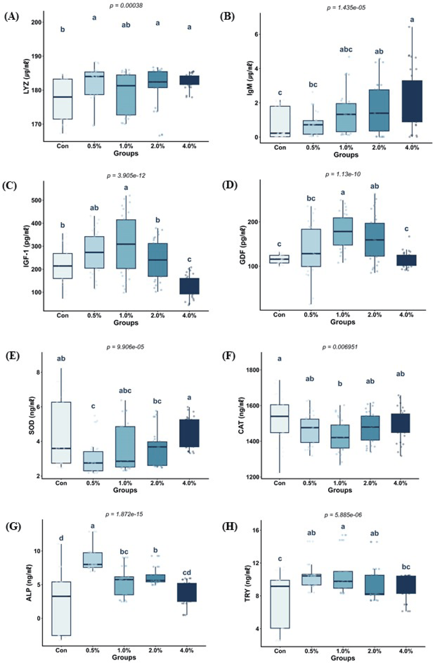
In addition, dietary supplementation with the additive significantly enhanced growth-related indicators. In particular, plasma IGF-1 levels exhibited a quadratic pattern, peaking at the 1.0% inclusion level (301.66 ± 41.88 pg/mL) and decreasing significantly at the 4.0% level (120.23 ± 74.54 pg/mL). Similarly, plasma GDF10 levels peaked in the 1.0% group (174.18 ± 60.89 pg/mL) and decreased in the 4.0% group, exhibiting a quadratic pattern.
Plasma SOD levels were significantly higher in the 4.0% group (4.80 ± 0.94 ng/mL) than in the 0.5% (3.46 ± 1.18 ng/mL) and 2.0% (3.90 ± 1.97 ng/mL) groups; however, there were no significant differences among the other groups. In contrast, CAT levels were significantly lower in the 1.0% group (1,429.88 ± 73.65 ng/mL) than in the control group (1,551.61 ± 116.98 ng/mL).
Furthermore, dietary supplementation with the additive significantly modulated intestinal digestive physiology. In the anterior intestine, ALP levels were significantly higher in the 0.5% group (8.86 ± 1.79 ng/mL) than in the other experimental groups, including the control group (4.97 ± 3.56 ng/mL). Similarly, TRY levels were significantly higher in the 0.5% (10.64 ± 1.79 ng/mL) and 1.0% (10.23 ± 2.17 ng/mL) groups than in the control group (7.33 ± 3.36 ng/mL).
To explore the mechanisms underlying the physiological changes, we examined the expression of specific genes in the intestine (Fig. 2). Significant differences in ApoAIV expression were observed in the anterior intestine (p < 0.0001). Dietary supplementation with the additive at 0.5% level significantly upregulated ApoAIV mRNA expression by 7.59-fold compared with that in the control group. Although not statistically significant, supplementation at other levels (1.0%, 2.0%, and 4.0%) increased ApoAIV mRNA expression by 0.43–2.64-fold compared with that in the control group. In contrast, dietary supplementation with the additive at 4.0% level significantly downregulated SOD and CAT mRNA expression by 0.37- and 0.16-fold, respectively, compared with that in the control and 0.5% groups (p < 0.0001), indicating that high inclusion levels of the additive (4.0%) may suppress antioxidant enzymes.
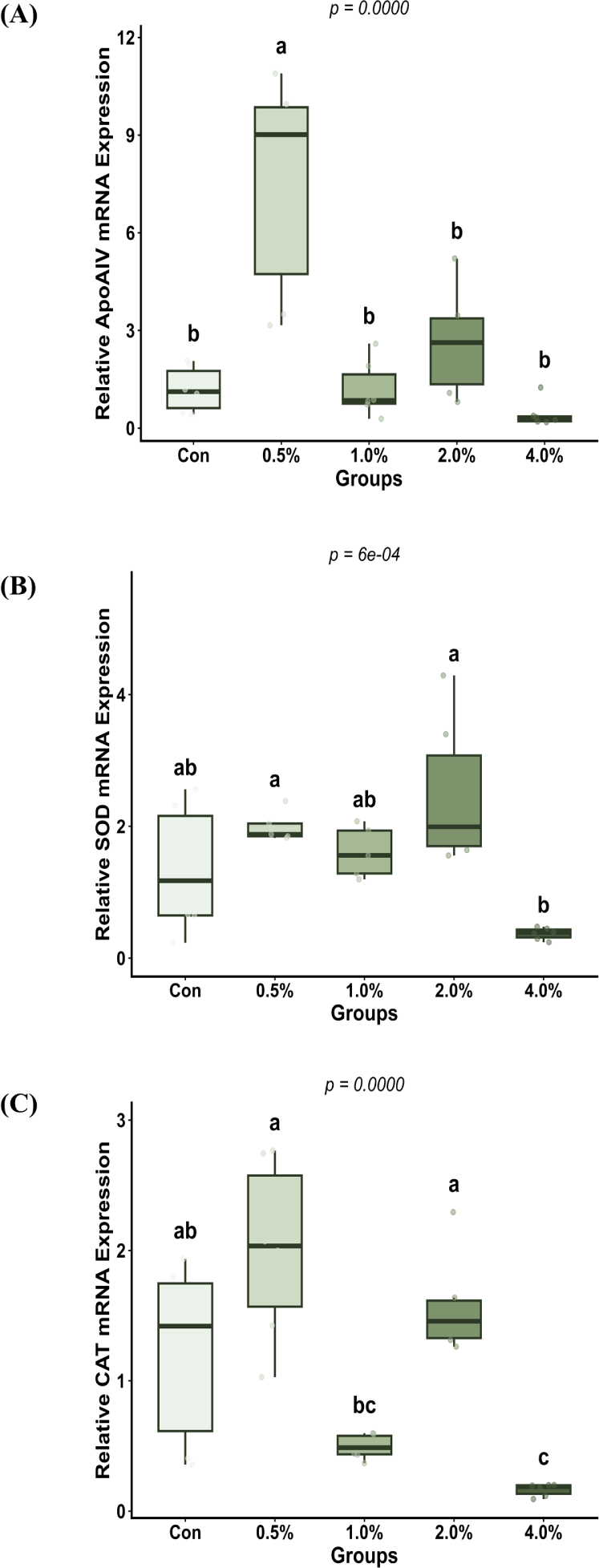
Table 4 shows the effects of the additive on intestinal morphology in fish. Dietary supplementation with the additive significantly influenced the number of goblet cells in the mid intestine (p = 0.014), with the 2.0% group exhibiting the highest increase (4,759.66 ± 405.76 cells), significantly outperforming the 1.0% and 4.0% groups. In contrast, no significant differences were observed in goblet cell numbers in the anterior or posterior segments. In addition, the 2.0% supplementation group exhibited significantly longer villi in the mid (1,539.73 ± 73.79 µm) and posterior (1,460.41 ± 56.27 µm) intestine than those in the 0 (control), 1.0, and 4.0% groups (p < 0.05). Collectively, these results indicate that a 2.0% inclusion level is most effective for enhancing the structural integrity and absorptive surface area of the intestine of olive flounder.
Discussion
Our study demonstrated that dietary supplementation with graded levels of a multifunctional bioactive additive containing glyconutrients, β-glucan, enzymes, and probiotics elicited differential responses in growth, immune function, digestive physiology, antioxidant-related responses, and intestinal morphology in olive flounder. Growth performance was most favorable in the 1.0% supplementation group, whereas intestinal histological parameters showed improvements in the 2.0% group. In contrast, the 4.0% group showed the highest plasma ALT and AST levels despite increases in some immune-related parameters. Overall, these findings indicate that the effects of this additive are not confined to a single biological endpoint but are mediated through multiple physiological pathways associated with growth, immunity, and intestinal health, with the overall response depending on the dietary inclusion level.
Although there were no significant differences in FCR and PER among the groups, fish in the 1.0% group exhibited the highest FBW, WG, and SGR, suggesting that the growth-promoting effect of the additive may not be attributable solely to improved feed efficiency, but rather to the combined influence of enhanced digestive and absorptive responses and improved intestinal conditions. Previous studies in olive flounder have shown that dietary supplementation with a synbiotic containing Bacillus sp. SJ-10 and β-glucooligosaccharide enhances WG and SGR and that supplementation with heat-killed Bacillus sp. SJ-10 or host-associated low-temperature probiotics improves growth performance (Hasan et al., 2018; Hasan et al., 2019a; Lee et al., 2024b). Similarly, other functional feed additives have been shown to improve growth performance in olive flounder, indicating that such additives can consistently contribute to growth enhancement in this species (Choi et al., 2023; Gunathilaka et al., 2022). Accordingly, the superior growth response observed in the 1.0% group confirms that this multifunctional bioactive feed additive can exert growth-promoting effects at an appropriate dietary level.
In terms of immune responses, IgM levels increased with increasing concentrations of the additive. In contrast, LYZ levels remained relatively stable among the treatments, suggesting that the multifunctional bioactive feed additive may have selectively modulated humoral and non-specific immune responses. Li et al. (2015) reported that dietary supplementation with 0.5% or 1.0% chitooligosaccharide significantly increased serum LYZ activity and other non-specific immune parameters in olive flounder and improved survival after pathogen challenge. Similarly, dietary supplementation of a low-fishmeal diet with multi-strain probiotics improved LYZ activity and antioxidant enzyme responses in olive flounder (Niu et al., 2019). In addition, a multifunctional immunostimulatory supplement enhanced LYZ activity and immunoglobulin-related responses (Medagoda et al., 2023). A recent study revealed that supplementation with probiotic- or yeast-derived additives enhanced LYZ activity in olive flounder compared with that in the control group (Choi et al., 2023). Collectively, the increase in IgM levels, together with the relatively stable lysozyme levels among treatments, suggests that the additive may have selectively modulated humoral immune responses, particularly through the combined actions of β-glucan and probiotics. However, these immune enhancements and growth and physiological parameters should be considered when determining the optimal inclusion level.
In terms of digestive physiology, supplementation with low-to-moderate levels (0.5%–1.0%) of the additive improved ALP and trypsin activities. In addition, the additive affected ApoAIV mRNA expression in the anterior intestine. Choi et al. (2023) reported that several functional feed additives significantly increased trypsin activity in olive flounder. Similarly, Lee et al. (2024b) suggested that host-associated probiotics could improve growth performance and digestion-related responses in this species. Overall, these results indicate that the changes in ALP and trypsin activity indicate that the additive influences intestinal digestive function or nutrient processing capacity. Given that direct digestibility indices, such as apparent digestibility coefficients, were not measured, the results should be interpreted cautiously.
Histological analysis revealed that supplementation with the additive (2.0%) increased the number of goblet cells in the mid intestine and improved villus length in the anterior, mid, and posterior intestine. Medagoda et al. (2023) reported that dietary supplementation with a mixture of nucleotides, β-glucan, and vitamins C and E significantly increased villus height and goblet cell count in olive flounder. In addition, Back et al. (2020) found that supplementation with heat-killed Bacillus improved intestinal fold density and intestinal wall thickness. Moreover, probiotic supplementation significantly increased intestinal villus length in olive flounder (Choi et al., 2025). Given that villus development is generally associated with an expanded absorptive surface area and that goblet cell abundance is related to reinforcement of the mucosal defense barrier, the histological improvements observed in the 2.0% group suggest that the additive enhanced the structural stability and functional integrity of the intestinal mucosa in olive flounder.
An important finding of this study was that the optimal dietary level for growth did not coincide with that for intestinal histology. Growth-related parameters were most favorable in the 1.0% group, whereas intestinal histological parameters were most clearly improved in the 2.0% group. This discrepancy suggests that the effects of multifunctional bioactive feed additives are mediated through the integrated action of growth-related endocrine responses, immune modulation, digestive function, and intestinal mucosal responses. Back et al. (2020) reported concurrent changes in IGF-I, LYZ, and intestinal morphological indices following probiotic supplementation. Choi et al. (2023) reported that growth, antioxidant capacity, digestive enzymes, and histological indices did not show similar trends in response to the additive. Overall, these results suggest that the optimal inclusion levels for improved growth and intestinal health are 1.0 and 2.0%, respectively.
In the context of antioxidant and plasma biochemical parameters, a high inclusion level (4.0%) requires more cautious application. In the present study, the 4.0% group showed relatively high values for some immune-related parameters, such as IgM, and significantly higher plasma SOD levels than the 0.5% group. In addition, the 4.0% group exhibited the highest plasma AST and ALT levels. Moreover, the high inclusion group did not show superior CAT and SOD mRNA expression compared with the low-to-moderate supplementation groups. Although SOD, LYZ, and blood biochemical parameters may improve depending on the type and level of functional additive in olive flounder, the responses are not always consistent among treatments (Choi et al., 2023; Hasan et al., 2019a; Niu et al., 2019). In addition, a recent study on the effects of probiotics in olive flounder reported treatment-dependent differences in ALT among dietary groups (Choi et al., 2025). Accordingly, the 4.0% treatment in the present study should not be interpreted as overtly toxic; rather, it may indicate that physiological benefits do not continue to increase proportionally beyond the appropriate supplementation range, and that some degree of metabolic burden may accompany excessive inclusion.
Proximate analysis revealed variations in the composition of the experimental diets among the treatments. As shown in Table 1, crude protein, crude lipid, and ash contents were not fully consistent across the dietary groups, limiting the extent to which all observed responses could be attributed solely to the additive. However, the positive responses observed in growth, immunity, digestive enzyme activity, and intestinal morphology at low-to-moderate inclusion levels suggest that this bioactive feed additive has potential as an effective dietary supplement for olive flounder. Overall, the practical dietary inclusion range of the additive is 1.0%–2.0%, with 1.0% being more favorable for growth improvement and 2.0% being more effective for enhancing intestinal health.
Because the additive used in this study was a mixture of glyconutrients, β-glucan, enzymes, and probiotics, the present experimental design does not allow us to distinguish the effects of individual components from possible synergistic interactions among them. Therefore, the observed improvements in growth, immunity, digestive physiology, and intestinal morphology should be interpreted as the overall response to the combined additive rather than as the effect of a specific single component. Further studies comparing individual components and their combinations, together with analyses of intestinal microbiota composition, direct digestibility indices, and tissue-specific immune-related gene responses, are necessary to clarify the underlying mechanisms.
Conclusion
Dietary supplementation with a multifunctional bioactive feed additive composed of glyconutrients, β-glucan, enzymes, and probiotics positively influences growth performance, immune responses, digestive physiology, and intestinal morphology in olive flounder. Among the tested levels, 1.0% supplementation was the most effective for improving growth performance, whereas 2.0% supplementation induced the most pronounced improvements in intestinal histological parameters. In contrast, 4.0% supplementation provided limited additional benefits and was associated with elevated AST and ALT levels, indicating that excessive supplementation may not be advantageous. Collectively, these findings suggest that the most practical inclusion range for this multifunctional bioactive feed additive in olive flounder diets is 1.0%–2.0%, depending on whether growth promotion or intestinal health is the primary target.